Traditional Biopsy Techniques vs. Tissue Microarray

Biopsies are critical for accurate diagnosis and treatment. The chronic shortage of cancerous tissue available for analysis is a difficult obstacle to overcome in the field of oncology. Traditional biopsies typically only include one case per slide, which is oftentimes not enough tissue to complete a thorough analysis. Tissue microarrays (TMAs) are used to analyze gene expression in numerous pathological samples on one single slide. 

Yale Pathology details the differences between conventional biopsies and tissue microarray services, highlighting the benefits of TMAs in both clinical and laboratory research settings. 

Conventional Biopsy 

  • One case per slide 

  • Requires many batches for a single study 

  • Requires a large amount of reagent

  • Subject to differential antigen retrieval

  • Very expensive and slow for large cohorts

  • 150 sections of one case yield only 150 assays 

Tissue Microarray

  • 50-500+ cases per slide 

  • An entire cohort can be done in a single experiment

  • Requires less than 1ml total reagent volume for the entire cohort

  • Not subject to differential antigen retrieval

  • Very rapid results, even when dealing with large cohorts

  • 150 sections can have over 500 cases, yielding up to 75,000 assays. 

Traditional Biopsy

A traditional biopsy is expensive and slow for large cohorts. They can only utilize one case per slide and require many batches for a single study, meaning it takes more resources and time to analyze the tissue available. Typically, medical professionals utilize needle biopsies at the time of diagnosis. A small amount of the tumor is removed, however, the needle typically does not remove enough tissue for a definitive diagnosis. In the field of oncology, a faster diagnosis means the patient can begin treatment sooner. 

Why Use Tissue Microarrays? 

Tissue microarrays are designed to study scarce tissue material to the fullest extent. Researchers can test hundreds of tissue samples with the antibodies of their choice, all at once. This reduces the amount of equipment, time, and space needed to study hundreds of tissue samples. Our customized tissue microarrays can be utilized to substantiate the importance of bio-targets in the advancement of diagnostics, therapeutics, and in considering new protein markers and genes.

How Does it Work? 

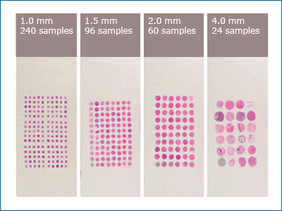
Currently, most tissue microarrays consist of paraffin-embedded tissues that are arranged into a paraffin block. The cancerous tissue is extracted from the subject by pricking the solid tissue with a circular needle and then sectioned onto the slide. The tissue is then preserved in the paraffin-embedded block. The needles can be sized around 0.6 mm to 4.0 mm, depending on how much tissue is available. The smaller the needle size, the more tissue samples can be collected. 

Comparison-of-tissue-microarray-samples.gif

Tissue microarray is proving to be an incredibly helpful technology in the field of science by increasing productivity, optimizing resources, and providing more accurate results. 

Personalized Tissue Microarray Services

Tissue microarray is the future of diagnostics in oncology. We are proud to offer customized tissue microarrays. Our geneticists provide customized tissue microarrays for target definition or biomarker investigation. Our donors are carefully chosen. We utilize a massive biorepository of aliquots that allows for quick access and error-free shipments. Our biorepository services are IRB compliant and adhere to the highest industry standards, ensuring the best experience possible. 

Purchase Your Tissue Microarray Now

Resources: 

FFPE Use in Next-Generation Sequencing

Overview

They are finding NGS next-generation sequencing analytical methods on DNA derived from FFPE clinical material is proving to be a powerful component in both oncological research and clinical diagnostics. Fresh- or snap-frozen samples are an excellent source for DNA, but they’re costly to collect and maintain and therefore have limited availability. This makes large scale retrospective studies of tumor-related cancers problematic, thus providing an open door for FFPE. Such studies typically give information regarding possible therapeutic targets as well as crucial prognostic information.  

Although there are many valid questions regarding the viability of FFPE, several studies have established that NGS can be performed using DNA from FFPE tissue successfully. The accuracy and reproducibility of such analyses, as well as their robustness to the bio-molecular quality of the samples used, is still the subject of many ongoing clinical studies. 

Comparative studies find that reasonable consistency of gene expression quantified from FFPE and FF specimens likely attributed the oligonucleotide probes measure expression being located at several positions across a gene. In addition to mRNA transcript quantification, microarray technology has been adapted to measure DNA copy number, single nucleotide polymorphisms (SNPs), and DNA methylation. 

Developing a Sequence Library from FFPE Samples

The preparation of the sequencing library is the first step in the NGS process. Many research outfits deploy different approaches in preparing a sequencing library. The variables in set-ups depend much upon the sequencing platform and the planned analysis (whole-genome sequencing, whole-exome sequencing, targeted DNA sequencing, whole-transcriptome sequencing, targeted RNA sequencing, ChIP-seq, RIP-seq, epigenetic studies.  

A sequencing library can be made by starting from genomic DNA or RNA. The workflow for the preparation of a DNA sequencing library consists of addressing the following areas:

  • Fragmentation and Sizing 

  • Attachment of the Adapters

  • Quantification 

The Benefits of Using FFPE Samples for Sequencing

Formalin fixed-paraffin-embedded (FFPE) tissue processing remains the most economical approach for long-term tissue specimen storage. Therefore, the potential to use FFPE preserved specimens for high throughput genomic applications to FFPE specimens is an active point of research to determine how accurate the analytical method is in practice. 

Clinical studies reveal certain advantages of working with FFPE versus Frozen in sequencing applications: 

Challenges with RNA FFPE samples

 The use of formalin-fixed paraffin-embedded (FFPE) samples offers unique challenges:

  • Acquiring high-quality sequencing data due to the wide distribution of sample quality

  • Differences in formalin fixation methods

  • Differences in storage conditions

  • Age of FFPE samples which may lead to cross-linked and/or degraded nucleic acid and inconsistent extraction yield

  • Insufficient DNA integrity or the presence of PCR inhibitors can lead to low read depth and allele dropout

  • Usage of FFPE specimens for ChIP-seq difficulties due to limited isolation of soluble DNA-protein complexes altered by excessive chemical cross-linking during the formalin-fixation process 

Best Practices for Using FFPE in Analytical Sequencing 

Because of the noted challenges using FFPE for various sequencing technology, the FDA weighs in harvesting methods and guidelines to help ensure a viable product. The National Cancer Institute (NCI) has published evidence-based best practices for FFPE DNA extraction, and the European Committee for Standardization Technical Specifications have also published a reference to the pre-analytical phase of FFPE sampled DNA.  

Tissue Procurement

The location of the biopsy and the amount of tissue harvested are of specific importance. These factors can influence the prediction of copy number variations. Despite the recent technical advances of NGS, tissue sample quality, and large amounts of DNA required often limit the sequencing process. 

Although it is essential for pathologists to understand the amount of tissue needed, it is likely even more important for the radiologist or surgeon to perform the procedure to acquire the tissue to understand. This makes communicating about the specific tissue requirements between the clinician and pathologist before the tissue harvest surgery an essential part of the success process. 

*The minimum quantity of DNA requested by most sequencing laboratories is between 50 and 200 ng. 

Comparable Success in Various Sequencing Methodologies Using FFPE vs. FF

Since miRNAs are more stable than RNA molecules, HTS is quite promising for quantifying miRNA profiles from FFPE specimens. These studies have found that miRNA-seq data generated from FFPE specimens have a similar number of total reads with just a slightly shorter average read length after trimming for adapter sequences.

DNA-seq has been modified to measure global DNA methylation patterns that are almost identical to methylation arrays using bisulfite treatment of DNA. Although less popular than DNA and RNA-seq, the use of FFPE in bisulfite sequencing has demonstrated a viable result. 

Conclusion

Multiple studies cite supportive data that accurate SNV can be identified from DNA-seq data from FFPE specimens. Despite the apparent concerns of FFPE specimen quality, it is widely accepted that paired with DNA-seq technology, it still holds value as it could provide the necessary data in order to fuel new therapeutic discoveries.

For more information on how Geneticist can help you procure large quantities of FFPE samples for your advanced sequencing project, please contact us. 

Biobanks and Tissue Microarrays: One of the Biggest Assets to Personalized Medicine and Clinical Research 

Overview

Human research biobanks are explicitly designed to act as a resource for high-quality biological materials, including Tissue Microarrays (TMAs). Tissue Microarrays, FFPE, and other related tissue samples are associated with the collection of clinical/biological data and organized for sharing among research scientists. In the recent decade, research biobanking structures are increasing in demand due to the innovation in technology and the vital role they are making in clinical research.

Quality biobanks are of tremendous value to the research community as they can offer specialized storage and transport conditions for a significant number of archived tissues in addition to having extensive tissue procurement and preservation services. Molecular tools for tissue profiling, such as expression microarrays, generally require the collection of fresh frozen tissues as sources of high-quality RNA. The fragile nature of RNA holds many concerns for scientists. Partnering with a reputable biorepository is essential to managing logistics and GMP protocols. 

Tissue Microarrays TMAs as a Research Tool

Clinical Tissue Microarray is a standard research tool used primarily for high-throughput molecular analysis of a variety of tissue types, including multi tumor microarray samplesThis analytical process is helping identify new diagnostic and prognostic markers and targets in many important health conditions such as tumor progression. TMA applications include use as a prognostic and research tool that assists in the investigation of morphology, protein and gene expression and/or chromosomal anomalies. Tissue microarrays for discovery and nonclinical work are usually either formalin-fixed paraffin-embedded or frozen TMAs. 

Tissue microarrays are composed of small-tissue cores as small as 0.6 mm in diameter, from regions of interest in paraffin-embedded tissues. These tissue cores are obtained using hollow needles of set diameters inserted into paraffin blocks. These cores are then transferred into slots in a recipient paraffin block in a precisely spaced array pattern. 

 Sections from the array block are cut using a microtome, mounted on a microscope slide, and analyzed by a variety of assay and staining techniques including IHC, ISH, FISH, TUNEL analysis, in situ PCR, and cDNA hybridization aided by laser-capture microdissection (LCM). Each microarray block can be cut into 50–1000 sections able to be used in various analytical processes, making it a cost-effective research vehicle.  

Biobanks Customize and Construct Tissue Microarrays (TMAs) for Use in Various Applications 

Tissue Microarray technology is based on nucleic acid hybridization between target miRNA molecules and their corresponding complementary components. TMAs are generally used to conduct a genome-wide analysis of miRNA expression of regular and/or disease samples, including cancer, and to distinguish expression signatures used in the scope of determining a diagnosis or designing a therapeutic intervention.

Tissue Microarrays (TMAs) in Translational Biomarker Research

Translational medicine (TM) is the emerging discipline involving the translation of laboratory findings into the design and implementation of early-stage clinical trials. The findings also help contribute to the drug discovery process. Translational medicine focuses on translating preclinical data from in vivo, in vitro, and in silico research into the clinic to help design trials, determine methods, and choose the biomarkers. With innovative analytical processes emerging at a lightning-fast rate, the use of TMA’s to help visualize biomarkers in neuroscience and immunological, inflammatory, and metabolic diseases are changing the way we explore treatment efforts as it relates to personalized medicine. Once the data is obtained, the pharmacodynamic biomarkers can then be applied to specific patient therapies and personalized medicine applications. 

Tissue Microarrays as a Diagnostic Tool in Personalized Medicine

TMA analytics assists in clinical-follow up and is used for providing personalized medicine, especially in cases of identifying histological changes. Personalized medicine seeks to identify individuals who will receive the most clinical benefit and least harm from a specific treatment. This is achieved by targeting genetic or other targets associated with their disease made possible by TMA, which rapidly analyzes and helps to identify with the biomarkers. Researchers can then organize and apply treatment guidelines based upon documented archival tissues and the corresponding study data.

Tissue Microarrays for Cancer Research

TMA technology is one of the hallmark methodologies used in the scope of oncological research. The medical community recognizes the value in quickly identifying changes in tumor activity and also the ability to store histological tissue safely for future use. TMA advanced staining techniques allow for advanced study of molecular behavior, which helps oncologists gain more insight on how to develop drugs that are more adept at treating cancer at various stages. v 

Conclusion

The various uses of TMA technology are exponential. Paired with quality biobanking services, tissue microarray as an analytical tool is paving a new direction for industries such as genomics, chemical engineering, and regenerative medicine. For details on customized biospecimen and tissue products such as FFPE, frozen tissue, and customized TMA’s, please contact us for more information. 

References:

https://www.ncbi.nlm.nih.gov/pmc/articles/PMC5963141/

https://www.sciencedirect.com/science/article/pii/B9780443069017500158

https://www.sciencedirect.com/science/article/abs/pii/S2307502314000320

https://www.tandfonline.com/doi/full/10.1080/14737159.2018.1522252

https://pubmed.ncbi.nlm.nih.gov/15094604-small-but-high-throughput-how-tissue-microarrays-became-a-favorite-tool-for-pathologists-and-scientists/

https://www.nature.com/articles/3780204

https://www.nature.com/articles/3700372

http://www.appliedclinicaltrialsonline.com/translational-medicine-and-biomarkers?pageID=2


What Are Clinical Trial Materials (CTM) Logistics and Why Are They Important?

Overview: What are CTM Logistics?

CTM Logistics is a quick reference to Clinical Trial Materials management services. CTM related management includes many moving parts and casts a wide net over the total breath of clinical management in general. In many ways, CTM logistics plays a crucial role in respect to meeting the needs of clinical environments. 

It goes without saying the priority is making sure specimens are getting to where they need to be with the integrity of the clinical product held intact. Protecting the viability is the most important thing to manage when it comes to implementing protocols for tissue transportation. Increasing the awareness of CTM details that require an additional layer of focus helps everyone involved become more solution minded.

An Open Look at What Makes the CTM Logistics Process Effective and Cost Productive

Meeting Volume Demands: This translates into being able to predict product demand and availability for the number of potential patients recruited for a clinical trial.

Global Management: There is much to think about when shipping overseas. Seasoned CTM managers need to be able to anticipate dealing with import/export restrictions and potential delays resulting in possible compromised environment and housing needed to optimize samples.

Labeling Challenges: Most companies do have some sort of data management in place. However, the failure comes into meeting the advance systems that allow for error-free end-to-end logistics. These systems address verification, validation, query, tracking, and coding for diseases or medications.

Training and Performance: Training is a game-changer and it extends to anyone who is in contact with transportation, processing, or handling of any type of biospecimen. Figuring out how to effectively manage a biorepository and meet the supply-chain demands is largely dependent upon training. Quality biorepository services plan for worst-case scenarios, preparation in ways that meet the GMP standards, and also maintaining best practices and SOP’s.

Biorepositories: What Role They Play in CTM Management 

At Geneticist, we place our emphasis on end-to-end logistical approach for all areas of clinical handling. We make sure we address every possible thing that might affect what really matters – and that is patient outcomes. As a major partner for most research endeavors, we understand how vital clinical specimen materials are to the overall big picture. Interestingly, this big picture can become very blurry when faulty handling measures are not overcome. 

Our role: What Geneticist does differently to help your project flow smoother. 

  • We focus upon the notification process by implementation of sophisticated technology and communication services. 

  • Our approach means placing our end-users as priorities and active within our communication loops. We integrate these people into the entire scope of the logistics process for seamless CTM strategizing and implementation.

  • We focus on standards and safe handling. The goal is to provide the highest standards available to protect the biospecimens from start to finish. 

A quote below from a respected market-leader helps drive home the fact the clinical trial platform has changed and is growing exponentially. CTM providers need to understand where we are and where we are going as far as being able to answer this big upswing.

“In the past 10 years and especially in the past five, the complexity of running clinical trials has grown enormously,” says Robert Pizzie, president of a newly formed consulting organization, Brizzey LLC (www.brizzey.com), expressly for clinical trial supply management.”

Global CTM Logistics: How to Service Hard to Reach Locations

In today’s current pharmaceutical landscape, there is something of an upswing going on and the need to focus on storage locations is a big deal. The market is driving the need to meet large capacities and heavy shipment volumes and frequencies. For large CTM outfits, this might be figuring out how to expand out to far-reaching places such as Latin America, Europe, and even Australia and creating what might be referred to as a global CTM network. This also includes in-country delicate transportation services where temperature-controlled vehicles will have to be provided and could the cause for potential delays. i 

CTM Logistics and Fragile Biologics

Investigational Medicinal Products (IMP’s) must be manufactured, stored, and distributed in accordance with the Good Manufacturing Practices, while also incorporating labeling and packaging that correspond with the appropriate clinical protocol. With sensitive biologics, there must also be special attention paid to optimizing the health of the specimen. This may include temperature controls, limited time frames, and specific housing environments or accommodation.

Conclusion

Clinical trials and related research are stretching beyond U.S. borders and also encompasses a strong global network. This indicates a significant logistical presence for anyone who is involved in the management of CTM’s. What we know is that as a full-service biorepository that we have to gain insight as to what our end users need in the way of fulfilling the demand in such a way that everyone benefits. With this in mind, we welcome strategic partnerships and will lend our expertise in any CTM logistics arena. Efficiency paired along with a strong service commitment is what will bring momentum to this long-neglected area.

References

  1. Expediting Clinical Trials with a Global Transportation Footprint and Clinical Packaging: https://www.pharmasalmanac.com/articles/expediting-clinical-trials-with-a-global-transportation-footprint-and-clinical-packaging

  2. FDA Resource for Best Practices Manual Update: http://www.aaei.org/wp-content/uploads/2015/12/FDA-Best-Practices-Manual-Update-FINAL-Sept-2015.pdf

  3.  Educational Resource for SOPs: https://biospecimens.cancer.gov/resources/sops/

What is the Tissue Procurement Process? 

The standard definition for tissue procurement is the administration procedure of acquiring tissues or organs for transplantation through various programs, systems, and organizations. Organ harvesting or tissue harvesting typically involves a surgical procedure and is especially relevant in the areas of molecular diagnostics. 

Why is Tissue Procurement Important? 

Tissue procurement is designed to help meet the ever-increasing demands indicated by a growing number of clinical researchers. Biospecimens used for analytical processes can come from diseased or non-diseased surgical, PMI, or autopsy donors. The applications prove to be invaluable in the field of molecular diagnostics. It is within this regard that we are seeing human tissues and cell -culture help to redefine how we solve medical dilemmas such as advanced disease and more effective treatment modalities. 

Understanding cell mechanisms of action prove to be one of our most useful advantages due to a modern shift within the biotech community. This also includes advancements in the preservation and related services helped in part by the strategic partnering with biorepositories to oversee tissue procurement procedures and storage. There are various methods of tissue preservation and a full range of biospecimens in which to order, complete with customizations. 

Geneticist is a full-service biorepository that offers:

  • Maintaining a comprehensive tissue database 

  • Providing histological staining and pathological review

  • Overseeing regulatory compliance

  • Coordinating clinical trials indicators and patient consent

Tissue Preservation Methodology

  • Frozen - Frozen tissue is snap-frozen in LN2 or frozen in cryo-embedding media such as OCT and stored at liquid nitrogen or -80° C temperatures until shipped.

  • Fresh - Fresh tissue can be minced or intact and placed in transport media of choice or saline and shipped for next day arrival.

  • Fixed - Provided in a fixative of choice or as paraffin-embedded blocks. Stained or unstained slides can also be provided from either paraffin blocks or as frozen sections. Fixed tissue can also be provided in a customized specified preservative, fresh or subsequently frozen.

  • Tissue Microarray slides are also available.

Tissue Procurement and Regulatory Considerations

The FDA has put in place a strong regulatory focus upon three general areas:

1) limiting the risk of transmission of communicable disease from donors to recipients

2) establishing manufacturing practices that minimize the risk of contamination

3) requiring an appropriate demonstration of safety and effectiveness for cells and tissues that present greater risks due to their processing or their use.

FDA regulates human cells or tissues intended for implantation, transplantation, infusion, or transfer into a human recipient. These are referred to as human cells, tissue, and cellular and tissue-based products. One of the main areas of research is related to cell culture. Their applications have expanded deep into the scope of drug discovery and other relevant therapeutic interventions. 

How Does Human Tissue Contribute to the Pharmaceutical Industry?

Helping to establish viable cell cultures is how we investigate the physiology and biochemistry of the cell. We can also determine the effect of various chemicals or drugs on specific cell types. This serves to examine the possibility for specific drug resistance which helps create more improved drug types and overall efficacy. 

The tissue procurement procedure most often occurs at the time of operation and it can be tested at the time by pathologists, or it can be preserved at which point the genetic material can be extracted and tests against gene-chips. The information gathered is used in both a diagnostic and prognostic capacity such as assessing likely clinical progression. 

A few of the major functionalities of using cell lines are so that we can use them for generating artificial tissues and to synthesize valuable biological compounds from large scale cell cultures such as in the case of developing therapeutic proteins. Using cell lines offer a few advantages in the way of providing consistency and reproducibility of results. It is important to note that cell structure can be altered and they also continue to grow. Cells are able to adapt to different culture environments by varying the activities of their enzymes. Special attention must be paid to successfully recreate the growth mediums of the original microenvironment.

Conclusion

Tissue procurement process is one of the many integral steps to the overall research and development cycle for the pharmaceutical landscape. The donor process has undergone some mainstream refinements to help manage safe specimen practices in an end-end structured format. Targeting avenues to help avoid tissue contaminants continues to be a primary focus. However, strict SOP adherence paired with effective regulatory controls contributes to efficiency in the overall tissue procurement process.  

References:

https://dctd.cancer.gov/NewsEvents/20190415_new_tissue_procurement.htm

https://science.howstuffworks.com/life/cellular-microscopic/cell.htm

https://www.ncbi.nlm.nih.gov/pubmed/17889099

https://www.fda.gov/vaccines-blood-biologics/tissue-tissue-products

https://www.ncbi.nlm.nih.gov/pmc/articles/PMC4089439/

https://www.ncbi.nlm.nih.gov/pmc/articles/PMC3341241/

https://www.lifegift.org/tissue-donation-process

https://www.ncbi.nlm.nih.gov/pmc/articles/PMC4724782/

The Availability and Type of FFPE Sample Collection Formats

The Availability and Type of FFPE Sample Collection Formats

Modern research efforts are fueled almost exclusively by the availability of human tissues. Tissue procurement is truly a fine-art or rather, a fine-science, however, you’d like to apply it. What is known about providing FFPE samples is that the research community demands the best quality assurance standards available. This is where partnering with a professional biorepository offers multiple layers of value to those within our industry platform. 

FFPE Sample Collection Formats 

Individual tissues: Diseased or normal from an individual donor

Four-tissue or quad combination sets: Fresh frozen, paraffin-embedded and formalin-fixed tumor and normal adjacent tissue specimens from one donor are available in combination sets

Matched pairs: 

FFPE and fresh frozen tumor and normal adjacent tissue samples from one patient.

FFPE diseased/tumor and metastatic tissue samples from a single donor.

*Most FFPE procured specimens are sized at 0.5 x 1 x 1 cm. This can vary significantly depending on the nature of the disease and tissue type. The fixation agent and the embedding media might also vary depending upon requested customizations.

*Most tumor tissues offer a 60% tumor content. 

FFPE Sample Collection Formats Used in Modern Applications 

It is easy to understand the significance placed upon usable tissue quality and diversity in assay preparation when you take into consideration the role it plays in medical advancements. Biomedical research requires a large quantity of tissues to analyze and characterize care strategies based upon what Is identified diagnostically through FFPE tissues or frozen specimens.

Geneticist moved in a progressive way to provide a vast array of customized tissue assays. In doing so, we are able to respond to the needs of researchers in a few different positive ways:

  1. The ability to offer an economic advantage for the researchers, as they can analyze multiple different tissues from different patients within the same assay. 

  2. It represents a global approach – availability and quantity matters. With customized tissue assays, we can arrange multiple assays in a way that represents a few different testing needs – this is especially true for advanced diseased research and best represented in quad tissue formatting. 

  3. Microarrays (TMA’s) are the preferred tool for large scale research efforts, as they allow the genome-wide measurement of transcription abundance.

There has been a huge shift in how tissue combination sets are used. Studies suggest that the consolidation of the four different tissue expression data sets can increase data quality which results in more meaningful interpretation.

FFPE sample collection formats are used in several different clinical applications such as:  

  • Oncology: (cancer four-tissue set, cancer serum/plasma, blood in special tubes)

  • Inflammatory Disease: (synovial tissue synovial fluid, matching blood, serum, plasma)

  • Neurological Evaluation: (postmortem brain tissue from patients with PD and AD)

  • Normal Individualized Tissues: (4 - 12 hours postmortem and surgical collection)

Quality Assurance 

Geneticist offers a full suite GMP certified specimen and/or tissue collection models that can be used in varying clinical capacities. Our team operates under the strictest SOP controls and we pair this methodology with IRB certified professionals for tissue procurement collection efforts. 

FFPE (formalin-fixed, paraffin-embedded) is a form of preservation and preparation for biopsy specimens that aids in the examination, experimental research, and diagnostic/drug development. More specifically, it normally involves a piece of harvested human tissue prepared by a certified medical pathologist that is submerged in 10% neutral-buffered formalin for 18 - 24 hours and embedded in IHC-grade paraffin. 

The harvest methods matter because the collection model is dependent upon viability. Storage and housing also matter, but the preservation of this type indicates FFPE is safely stored at room temperatures. However, all specimens are housed with specific adherence to GMP and other regulatory and laboratory best practices in mind. 

Addressing the Concern: Obtaining Viable FFPE and Frozen Tissues from a Biorepository

The operational procedures of the biorepository must be well-trained in order to match that which is provided to the investigator versus the overall diagnosis of the specimen via a surgical pathology report. Specimen mismatching occurs in many instances such as in the case of necrosis, or unsuspected tumor invasion of seemingly normal tissues in which the specimen is mistaken grossly for tumor.

During the course of what may be called a huge push in genomics and other related “omic” genres, doubts and concerned have surfaced. The goal of obtaining viable FFPE tissues are centered around reproducibility making the procurement and handling methods a critical element.  There are relevant studies which have raised eyebrows citing poor reliability of microarray data based upon incongruent preparation methods of varying TMA platforms.vii

The bottom line is there will always be factors to weigh in on such as TMA platform, tissue handling, RNA isolation method and hybridization procedure which have the potential to alter the results. We maintain at Geneticist that these factors are greatly minimized when the tissues are collected with a high level of acuity and applied to consistent levels of output. 

References:

  1. https://www.ncbi.nlm.nih.gov/pmc/articles/PMC3445033/

  2. https://www.ncbi.nlm.nih.gov/pubmed/21898218

  3. https://www.ncbi.nlm.nih.gov/pubmed/15542899

  4. https://www.ncbi.nlm.nih.gov/pmc/articles/PMC2664171/

  5. https://www.sciencedirect.com/science/article/pii/S0888754318300995

Biorepositories– The Most Valued Resource of Modern Research

Biorepositories– The Most Valued Resource of Modern Research

What is a Biorepository or Biobank?

The standard definition is a place that collects, stores, and distributes biospecimens to requested designations for research purposes. The truth is that most biorepositories serve a much greater field of services than what meets the eye. In scientific circles, they are a valuable asset to laboratory research and over 120 biobanks that are used worldwide. Biorepositories are so valued in fact, Time Magazine published an article citing that biobanks are among ten of the ideas slated to change the world. (1) (2)

The Primary Function of a Biorepository

The main functional component of the biorepository is to house various biologics. The types of specimens stored include blood, urine, tissue, stool, and not limited to human specimens, but can also be animal. Most of the human specimens have medical data stored in conjunction with them and are cataloged for future use. These biologic samples can be designated for private or public project partners such as vaccine manufacturers, regulatory agencies, molecular diagnostic companies, and oncologic research endeavors. (3)

Regulatory and Privacy Concerns

Biorepositories are not without a set of checks and balances. They aren’t allowed to operate under a set of closeted rules that can potentially interrupt public safety. It is important to note that biospecimens contained within a cGMP certified biorepository are under strict regulation from the government and they are required to adhere to the rules and certification standards that apply. The role of bioinformatics within the industry assists to regulate the flow of information as it applies to the large libraries of cataloged specimens. There is an ever-increasing burden to encrypt biospecimen collections with sophisticated security measures that protect patient privacy and also ensure accessibility. (4) (5)

Supporting the Demand of Special Research Projects

One of the main functions is housing large volumes of biologics. Biospecimens are considered precious to the world of drug discovery and development. Genetic material helps to develop novel breakthroughs in the world of advanced disease and, as such, are in high demand many clinical studies are the world. The number of registered clinical trials has increased significantly in recent years. As of August 2019, there were about 314,644 thousand clinical studies registered globally. (4) (5) (7)

Quality Control 

Capable of digitally managing the characterization of specific cultures, tissue types, and fluids, biorepositories are one of the most reliable sources for obtaining viable laboratory study mediums. Viability being the optimal word- maintaining the integrity of the specimen is paramount. Laboratory professionals run a tightly wound operation that oversees the maintenance of these biospecimens to help reduce errors in testing and quality. Management includes the complexities of the freeze-thaw cycles and understands how to effectively minimize contamination. The goal is to produce reproducible efficacy in a clinical test sample. The biorepository is essentially the most important element of transition between the harvest point of the biospecimen and the clinical endpoint of the intended study. (7) (8)

Diverse Range of Services

Cell and Tissue Banking 

  • Peripheral blood serum/plasma/mononuclear cells (PBMC)

  • Lamina propria mononuclear cells (LPMC)

  • Bone marrow cells/plasma

  • Lymph nodes

  • Human stool and isolated stool bacteria communities

  • Buccal swabs (Human mouth epithelial cells for DNA purposes)

  • Tumor Tissue

  • Pleural Fluid

  • Thymocytes

  • Splenocytes

  • Bronchoalveolar lavage fluid

  • Extracted DNA

  • FFPE/Frozen Tissues

Laboratory Services

  • Laser capture microdissection

  • Tissue microarray construction

  • IHC preparation

  • RNA, DNA isolation

  • Digital Imaging

  • FFPE or Frozen tissue preparations

  • Custom microarray construction

CRO Services

They offer clinical monitoring services such as acting as a site monitor for clinical research projects, develop customized SOP’s, or consult on special clinical investigations or investigational drug protocols. 

CTM Logistics

They specialize in arranging temperature-controlled delivery of biological samples and overseeing the collection, retrieval, and extraction processes. 

Modernizations in Biorepository Management

Even with the diverse range of services and capabilities of biorepositories such as Geneticist, in a modern technological landscape, more streamlining is required. We can look forward to the continued contributions in the form of better specimen traceability and management. As the growing amount of unique requests are needed with respect to creating customized arrays, the industry recognizes being able to highlight the unique origins and characterizations via small digitized footprints that are easily shared among genomic professionals. 

Modern laboratory science not only depends upon quality controls in the lab but also cloud-based sophisticated inventory management that pairs nicely with tracking and easy sharing. The adoption of a changing landscape is reflected in modern biorepository spaces. Many are equipped with high functioning, lighting quick lab computing interfaces with respects to temperature controls, biodata, easy accessibility, and permission controls, and ability to share with other biobanks or LIMS systems. (9) (11) (12)

References and Further Reading

1. [Online] http://content.time.com/time/specials/packages/article/0,28804,1884779_1884782_1884766,00.html.

2. [Online] https://www.ispecimen.com/blog/2018-will-big-year-biobanks/.

3. [Online] https://www.ncbi.nlm.nih.gov/pmc/articles/PMC6132819/.

4. [Online] https://www.ncbi.nlm.nih.gov/pmc/articles/PMC3038212/.

5. [Online] https://cancerworld.net/impact-factor/biorepositories-for-cancer-research-in-developing-countries/.

6. [Online] https://www.statista.com/statistics/732997/number-of-registered-clinical-studies-worldwide/.

7. [Online] https://www.ncbi.nlm.nih.gov/pubmed/26420610.

8. [Online] https://www.ncbi.nlm.nih.gov/pubmed/21745162.

9. [Online] https://www.ncbi.nlm.nih.gov/pubmed/1367056.

10. [Online] https://www.ncbi.nlm.nih.gov/pmc/articles/PMC6132819/.

11. [Online] https://www.ncbi.nlm.nih.gov/pubmed/26514206.

12. [Online] https://www.ncbi.nlm.nih.gov/pubmed/26418270.


Biospecimen Collection and Extraction Processes 

Biospecimen Collection and Extraction Processes 

Biospecimen collection and extraction processing is the step describing the way donor samples are collected from the donor-source, preserved, labeled, and then stored or delivered. Human biospecimens are usually obtained from a patient for a certain test or diagnostic procedure. In some cases, patients give advanced permission for more tissue to be taken than needed for the purposes of research. Consequently, it is not uncommon for biorepositories to store large quantities of frozen or FFPE tissues taken from donor patients and designated to different areas of research. (1)

Is There a Special Process of Retrieving Biospecimens? 

The receipt or retrieval of biospecimens is handled by dedicated laboratory professionals often referred to as specimen processors. The laboratory staff at Geneticist works under a strict set of standard operating procedures (SOPs) that are commonplace in our industry. We find that by incorporating SOP’s and appropriate safety methods and procedures, we are able to better protect the viability of the specimens housed in our biorepository. 

Specimen processing practices involves bringing into play appropriate handling guidelines set forth by laboratory professionals and regulatory agencies such as the CDC. The function of a biorepository is to facilitate the cycle the donor specimens have to travel through and arrive stable enough to be used in research or diagnostic capacities. Additionally, we do take into active note the requirements the IRB places on existing versus perspective samples and how it relates to clinical studies. (1) As a result, our biorepository maintains strict adherence throughout the entire cycle of specimens handling. (1) (4)

Is It Difficult to Avoid Specimen Contamination? 

Handling specimen donor samples does require a high level of acuity. A laboratory personnel member must be trained specifically to handle specimen collection and processing duties. Once the specimen is removed from its original environment, the risk of contamination is a real possibility. Hence, the process of specimen extraction is an important component in maintaining the integrity of the sample. 

Specimen collections can involve a variety of different scenarios, especially with varying types of tissue samples and biofluids such as: 

  • Plasma

  • Urine

  • Saliva

  • Stool

  • Whole Blood

  • Hair Follicles

A dedicated area or space is often delegated for the strict purpose of assisting the investigator to collect donor samples. Those samples then either go on to specified research designations or to other to long-term bio-storage. 

Factors that can alter the stability of a specimen sample include: 

  • Temperature changes

  • Faulty preservation standards

  • Toxic Exposure

  • Time – (Handling, Transport, Processing, Exposure)

  • Type of Anesthesia 

  • Field Contamination

Preanalytical Variables – Preparing Biospecimens for Use

Preparing the biospecimen for testing is a marriage of quality control and error-free methodology. Any variances with regards to the sample can affect the performance of the testing performance of the assay. Specimen handling is critical so as to avoid changes in the molecular structure of the sample. It is cited in a government article that as much as $28 Billion is spent each year on irreproducible research. This translates to inconsistencies in specimen management. (5) 

Consequently, much attention is paid in the areas of specimen handling time, storage temperatures, size, and type. There is a myriad of variables that contribute to the biospecimen process as a whole. Before a sample can be stored for use in other capacities, there is a series of processes in which a sample must undergo prior to the analytic process. 

What Are the Some of the Ways Human Biospecimens Are Used? 

Whether the tissue extracted is needed for diagnostic reasons or for storage, the practice of obtaining donor samples is the cornerstone of drug discovery and the study of advanced disease. Researchers commonly use FFPE samples, frozen tissues, and other biospecimens in various scientific modalities. The use of human tissues in clinical studies has contributions to just about every area of medicine, advanced disease, and oncologic studies. (6) (7)

FFPE is one most common types of biospecimen samples used in a specific modality referred to as IHC or immunohistochemistry. These IHC applications are used for studying advance disease characterizations and aspects in the fields of hematology, immunology, Alzheimer’s research, and comparative studies. (6)

Preparing an FFPE involves preserving the tissue in a formalin-formaldehyde derivative and then fixed onto a slide. The slides can represent the isolation of a protein, RNA, DNA, or rare tissue types on the same assay for comparative views and analytical purposes. The general nature of the FFPE is a preservation by which it is favored in many circles because of the longevity and versatility of sample use.

Frozen tissues also are represented well in research. They are used for examining cell structure, type, and mechanism of action. (9) It is important to note that all human tissues or specimens are somewhat volatile. In the scientific community, there are noted differences between the specific preferences of FFPE tissues or frozen tissues – especially in terms of their long-term storage capabilities. Geneticist provides both types and any requested customizations.

References and Further Reading:

1. [Online] https://bios.ucsf.edu/what-are-human-biospecimens.

2. [Online] https://irb.ucsf.edu/research-using-human-biological-specimens#common.

3. [Online] https://biospecimens.cancer.gov/bestpractices/to/bcpsrd.asp.

4. [Online] https://www.ncbi.nlm.nih.gov/pmc/articles/PMC3995361/.

5. [Online] https://www.ncbi.nlm.nih.gov/pmc/articles/PMC5073593/.

6. [Online] https://www.ncbi.nlm.nih.gov/pubmed/21347257.

7. [Online] https://www.cancer.org/treatment/understanding-your-diagnosis/tests/testing-biopsy-and-cytology-specimens-for-cancer/what-happens-to-specimens.html.

8. [Online] https://www.ncbi.nlm.nih.gov/pubmed/30113239.

9. [Online] https://www.nature.com/articles/srep39394.


What Are the Various Applications of Biomarkers? 

Biomarkers are used in various applications related to these three main areas:

  1. They help to assign a certain amount of predictability for certain illnesses.

  2. They can help to identify precursors for advanced diseases such as cancers or blood disorders.

  3. They play a major role in the drug discovery and development process

Clinical thought leaders actively engage the idea that biomarkers are the centerpiece for finding revolutionary discoveries in medicine and in pharmacologic endeavors. (1) Biomarkers are a crucial element to a majority of scientific research and medically necessary to determine the potentiality of disease and exposure. (2) As an integral component of the clinical trial process, biomarkers serve as a method to determine the efficacy or effect of many medical-related procedures and circumstances. Additionally, biomarkers are one of the valued measurements for the effects of toxic exposure. (3)

Biomarkers Are Essential to the Drug Discovery and Development Process

Biomarkers studies require the processing and storage of numerous biospecimen samples for the purpose of storing information and reducing research expenses. Biorepositories maintain the characterized samples and usher them to specified research purposes and clinical applications.  Biomarkers are multi-dimensional in terms of usage. The chief function that they offer a set of directional cues for researchers. The big scientific shift into genomics, bioinformatics, cytometry, and other related modalities is helping to find specific biomarkers that are associated with solving the riddles related to chronic disease. (4) (5)

Biomarker Classifications 

    • Exploratory Biomarkers - Tools used at the preclinical phase without evidence necessarily linking the biomarker to a designated clinical outcome in humans. 

    • Demonstration Biomarkers - This means the biomarker has been qualified (not validated) in terms of preclinical sensitivity and is linked with specific clinical or patient outcomes, but without having to be reproduced in the context of a clinical study. 

    • Characterization Biomarkers - demonstrates adequate preclinical sensitivity and specificity and are able to link to clinical outcomes. The progression of qualification leads to increased use of a biomarker in decision making regulatory applications.

Validating biomarkers

Some of the biggest challenges are to validate and characterize large quantities of bio-tissue samples to meet the demand. (6)This is where strategic partnerships with biorepositories such as Geneticist comes into full view. We process and house large quantities of biospecimens that are viable enough to move forward to the clinical trial process. The scientific community had some concerns about the slow progress of biomarker research due to low standards in specimen collection and handling paired with overall poor trial design. (7)

Biorepositories and Biomarker Processing

Geneticist is the gold standard when it comes to the logistics and technical complexities of biomarker validations. “Omic” platforms I.e. genomics, proteomics, etc., rely upon the ability to put into play large quantities of analytes in single customized assays – we provide this support to our research partners. We can prepare validated assays needed to facilitate the rapid generation of data needed to qualify the biomarker for reuse in another purpose. (6) (9)

Understanding the Differences Between Biomarkers and Clinical Endpoints

When trying to find the distinguishing aspects between biomarkers versus a clinical endpoint, we only need to evaluate their chief point of function to find the difference. The major function of a biomarker is to find a point of observation that best represents the existing clinical circumstance. Conversely, the major function of a clinical endpoint is to identify an outcome. (10) It can be a primary or secondary notation, and it most often addresses an answer to a research question. 

  • Biomarkers act as an objective or observed point of reference to identifying a quantifiable measurement. They do not take into consideration the patient’s perspective. (6) 

  • Clinical Endpoints represent a clinical outcome measured by a clinical trial and include a first-persons point of view or perspective. It answers questions: How did the patient feel? How did the patient function? Did the patient survive? For example, survival is considered to be the most optimal clinical endpoint of HIV related studies. (12)

Example: 

Patient-reported fever. Has elevated WBC. 

Will Tylenol resolve the fever? Tylenol was given at 3 pm. 

Endpoint answer: Fever ended at 5 pm. The patient feels better. The cessation of symptoms ended after 3 hours. (An endpoint serves as an answer to whether or not the Tylenol resolved the fever.)

Biomarkers: Elevated white blood cell count paired with fever is a biomarker thus implying an infection within the body. 

The Future of Biomarkers

Trending, is the application of exosomes in use in various clinical capacities. Exosomes are extracellular vesicles first described as much as 30 years ago in regards to cell-communication and disease transmission. They are considered by many to be one of the most valuable biomarkers because they contain a footprint for cell mechanism of action.  It is speculated that Exosomes will be best used as a diagnostic and prognostic tool for malignant tumors. Biorepositories can safely store exosomes for decades, as they tend to maintain their viability if stored under the proper conditions. (11) (12)

References and Further Reading

1. [Online] https://patientdaily.com/stories/510644132-medical-thought-leaders-call-for-action-on-biomarkers.

2. [Online] https://www.ncbi.nlm.nih.gov/pmc/articles/PMC534923/.

3. [Online] https://www.ncbi.nlm.nih.gov/pmc/articles/PMC534923/.

4. [Online] https://www.sciencedirect.com/science/article/pii/S0041008X05000864.

5. [Online] https://www.ncbi.nlm.nih.gov/pubmed/22523699.

6. [Online] https://www.ncbi.nlm.nih.gov/pmc/articles/PMC6037391/.

7. [Online] https://www.cell.com/trends/molecular-medicine/pdf/S1471-4914(12)00177-3.pdf.

8. [Online] https://www.europeanpharmaceuticalreview.com/article/4357/biomarkers-drug-discovery-development/.

9. [Online] https://www.cell.com/trends/molecular-medicine/pdf/S1471-4914(12)00177-3.pdf.

10. [Online] https://www.focr.org/clinical-trial-endpoints.

11. [Online] http://www.proofcentre.ca/resources/biomarker-solutions/what-are-biomarkers/.

12. [Online] https://www.focr.org/clinical-trial-endpoints.

13. [Online] https://www.ncbi.nlm.nih.gov/pmc/articles/PMC5502131/.

14. [Online] https://bmcbiol.biomedcentral.com/articles/10.1186/s12915-016-0268-z.


The Two Types of Bone Marrow

The Two Types of Bone Marrow

Bone Marrow Transplantation and Stem Cell Therapies are literally “the heart” of many clinical trials and research efforts. (1) Many people are struggling to survive and cope as they live with chronic diseases, blood disorders, and cancer. As a result, the science community is actively working with biorepositories to acquire quality bone marrow and stem cell samples needed for clinical research. (2)

What Exactly is Bone Marrow? 

Bone marrow is the soft, malleable tissue located in the center of the bone. It is comprised of connective tissues, blood vessels, and capillaries that are responsible for the production of different cell types that create and sustain life. Bone marrow is responsible for generating billions of new blood cells every day. There are two distinct types of bone marrow – yellow marrow and red morrow. On average, most adults have about 2.5kg of bone marrow, half of which consists of the red morrow type. 

Red Bone Marrow (Myeloid Tissue)

Red Bone Marrow is responsible for producing three primary cell types:

  • Red Blood Cells (RBCs) - Red blood cells are responsible for carrying oxygen throughout the body. It also carries carbon dioxide away from the tissue and back to the lungs to exhale. Red blood cells have a life span of about 120 days.

  • Platelets – Platelets are responsible for preventing excessive bleeding and assist the blood clotting process after injury. Platelets have a life span of about ten days.

  • White Blood Cells (WBCs) - White blood cells assist the body in fighting infection. White blood cells have a life span of only a few hours to a few days.

When we are born, our bodies contain all red bone marrow. Later, as we age, it becomes yellow bone marrow. It is found mainly in the medullary cavities (within the flattest bones) of the body: hip bone, breast, skull, vertebrae, ribs, shoulder blades, and in the cancellous material at the ends of the long femur bones and humerus. (3) 

Yellow Bone Marrow

Yellow Bone Marrow is called yellow because of its high concentration of fat cells. It is responsible for aiding in the storage of fats in cells called adipocytes. Yellow bone marrow is responsible for producing cartilage, fat, and bone. In cases of severe blood loss, the yellow bone marrow has the potential to turn back from yellow to red bone marrow. 

Bone Marrow Contains Two Types of Stem Cells

Bone marrow produces two specific types of stem cells, hemopoietic stem cells, and stromal stem cells. These are human cells with the ability to differentiate into other types of cells, making them the building blocks of functional life. Researchers discovered that stem cell-derived therapies are capable of correcting advanced disease, blood disorders, re-growing limbs, and regenerating failing organs. 

Hemopoietic Stem Cells (HSCs)

HSCs are the foundation cells for the production of red blood cells, AND it is capable of renewing itself. HSCs are essentially a multipurpose type cell, they can differentiate themselves, but it also has the power to mobilize away from the bone marrow into the main bloodstream and undergo apoptosis (self-destruction). 

 Mesenchymal (MSCs) or Stromal Stem Cells 

These are cells harvested from adult bone marrow, adipose tissues, neonatal tissues. They make up the connective tissues (the supportive tissues surrounding vital organs). The most common stromal cell types are fibroblasts and pericytes. The highlight of these types of stem cells is their ability to proliferate and their immunosuppression functionality. (4) 

Bone Marrow Diseases and Transplantation

Genetics and other factors can hinder the ability of the bone marrow to function properly. Abnormal functionality of the bone marrow’s ability to create stem cells results in bone marrow disease or disorders that require the need for transplantation and/or advanced treatment. (4) 

Bone marrow diseases are challenging to treat and are, in most cases, assigned a poor prognosis. This is where the bone marrow transplants come into practice. (4) However, one of the biggest challenges is to a bone marrow donor capable of matching the patient in need. (5) 

Did You Know? 

Only 30% of patients requiring a bone marrow transplant have a matching bone marrow donor within their family unit.

The two most important factors in matching a donor to a patient are 1) age and 2) ancestry. 

Bone Marrow Diseases:

    • Leukemia - a cancer of the blood where the bone marrow makes abnormal white blood cells

    • Aplastic Anemia – A condition in which the bone marrow does make a sufficient amount of red blood cells.

    • Myeloproliferative Neoplasms (MPN) - This is a cancerous condition that comes about when the body makes too many red or white blood cells and platelets

    • Lymphoma cancer– While not directly affecting the bone marrow, it involves the immune fighting cells o the immune system that resides in the bone marrow, spleen, and other parts of the body.

 Bone Marrow and Stem Cell Research 

Bone marrow and their stem cell producing abilities take center stage when it comes to new drug discoveries and their potential to regenerate and heal the body. Researchers use both types of bone marrow extensively for research purposes to help understand better their diverse mechanisms of actions. The future of bone marrow transplants and solving the mysteries of advanced diseases hinge upon finding ways of harnessing the cell differentiating power of stem cells. (7) 

Using a Biorepository for Long Term Storage for Bone Marrow and Stem Cell Products

In order to facilitate scientific research of bone marrow, the research community depends upon the long-term storage of bio-specimens, bio-fluids, stem cells, and bone marrow. Geneticist understands the sophisticated methods of cryopreservation to maintain the viability of stem cell products for the purpose of research. (7) Our laboratory professionals are also able to handle the logistics of getting the samples delivered in such a way that it doesn’t disturb the integrity of the bio-product. 

References and Further Reading

1. [Online] https://www.nature.com/bmt/.

2. [Online] https://biospecimens.cancer.gov/bestpractices/to/bcpsrd.asp.

3. [Online] https://www.sciencedaily.com/terms/bone_marrow.htm.

4. [Online] https://link.springer.com/article/10.1007/s00018-019-03125-1.

5. [Online] https://www.medicalnewstoday.com/articles/285666.php.

6. [Online] https://www.niddk.nih.gov/health-information/blood-diseases/aplastic-anemia-myelodysplastic-syndromes.

7. [Online] https://bethematch.org/support-the-cause/donate-bone-marrow/join-the-marrow-registry/likelihood-you-will-donate/.

8. [Online] https://publons.com/journal/27172/journal-of-bone-marrow-research.

9. [Online] http://www.hcpro.com/HOM-57532-3631/Rules-Regulations-Guidelines-for-stem-cell-storage-and-collection.html.


Tissue Microarrays: A Modernization of the Bio-Specimen Industry

Tissue Microarrays: A Modernization of the Bio-Specimen Industry

Background

Tissue Microarrays (TMA) are one of the premier methods of organizing specimens to facilitate diagnostic research. The TMA method, introduced by H. Battifora in 1986, is used extensively in the field of histology. (1) The creation of a tissue microarray serves as an avenue to analyze multiple specimens at the same time. (2)

One microarray or “tissue chip” contains many small representative tissue samples organized into one histological slide. A tissue microarray is comprised of a paraffin block produced by extracting cylindrical tissue cores from different paraffin donor blocks and re-embedding these into a singular block. A paraffin block can be housed at room temperature for years and hold up to 1000 tissue samples. (2)

TMA Applications 

Tissue biopsies are a staple in the medical field for both diagnostic and research purposes. Traditional tissue sample methods require a healthy amount of tissue and the process is both labor-intensive and time-consuming. From a pathological standpoint, TMA slides are a game-changer. The old way involved creating a new slide for each potential genetic marker for each individual patient or donor. (3)

With TMA, a whole slide approach can now be screened for several different biomarkers from the same patient – a complete thorough process towards understanding mechanisms of action. This is where using a quality biorepository is an integral part of major research objectives. Medical professionals have at their fingertips the ability to order customized tissue microarray samples. (5) Those samples can then be utilized in the scope of various analytical methodologies such as: 

  • IHC (Immunohistochemistry): The preferred analytical method for in-situ (place of origin) protein expressions in tissue samples

  • RNA: A popular technique that uses RNA sequencing for transcriptional profiling, editing, and analyzing gene expression. 

  • FISH (Fluorescence in situ hybridization): A technique that uses fluorescent probes that bind only to those parts of a nucleic acid sequence with a high degree of sequence complementarity. 

TMA Variations 

Tissue procurement is a scientific art form that has zero room for mistakes or interpretation. A quality biorepository will be able to process tissue specimens for a multitude of uses and analytical methods. SOP comes into strong play alongside integrity for the practice itself – biorepositories is a foundational element for all research activities. (6) The following are variations commonly used:

Formalin-Fixed Paraffin-Embedded Tissues (FFPE)

A tissue of preservation for biological specimens that aid in the examination and experimental research, and diagnostic development. Formalin is a formaldehyde derivative that helps preserve vital proteins and structural components within the tissue sample.

Frozen Samples

Frozen or Cryosection samples are often used in the context of oncological surgery as opposed to research. Although they are primarily used for rapid diagnostic contexts, frozen samples are used to view cell morphology, structure, and biochemistry disease, and mechanisms of action. The applications, pros, and cons are easily distinguished in laboratory science. 

Customizations

The chief benefit of working with Geneticist is that we tailor TMA samples unique to our clients. This helps scientists more efficiently analyze gene and protein expressions.

Why Do IRB Compliant TMA and FFPE Samples Matter? 

There are three types of research concerning specimens. Each type is governed by strict regulatory standards and federal mandates. The rules are in place to ensure specimen collection meets the ethical and safety standards set by many various governing bodies. The role of biorepositories crosses all regulatory frameworks and therefore requires the highest adherence level. The aspects that pertain to each are varied, but similar in the fact research activities and methods are subject to intense scrutiny throughout the following submission applications: drug discovery, new drug submissions, clinical trials, and modifications. (7) 

Failure to meet these standards can halt or slow the approval of anyone these processes resulting in significant losses of time and money. Hence; the importance of providing IRB compliant tissue specimens is the benefit of ordering samples from a biorepository with an excellent track record and relevant partnerships with research sponsors who are also held in high esteem. 

Research Specimen Types

  • Prospective Sampling: Samples have been approved through an IRB approved protocol for non-research purposes. 

  • Specimen Repositories: Tissue banks that have data associated with them i.e. demographic, behavioral.

The Role of IRB and Biorepositories

Biorepositories house thousands of specimens and samples of various kinds and they are required to have an approved IRB plan in place. The FDA and other regulatory agencies such as HIPAA, and  OPHS also maintain active involvement and rules of operation for tissue banking facilities. These regulating interventions are in place to oversee the collection, storage, and distribution of all the samples. (8)

Geneticist maintains all regulatory standards and our specimens and methodologies meet every compliance benchmarks. We also ensure the strictest controls and optimal storage solutions for all samples including biologics, blood serums, tissues, and other bio-components for the use of analytical research.

References:

1. [Online] https://www.ncbi.nlm.nih.gov/pmc/articles/PMC3599403/.

2. [Online] https://www.ncbi.nlm.nih.gov/pubmed/3525985.

3. [Online] https://www.ncbi.nlm.nih.gov/pmc/articles/PMC2813639/.

4. [Online] https://www.ncbi.nlm.nih.gov/pmc/articles/PMC2813639/.

5. [Online] https://www.ncbi.nlm.nih.gov/pmc/articles/PMC3599403/.

6. [Online] chrome-extension://oemmndcbldboiebfnladdacbdfmadadm/https://publications.iarc.fr/_publications/media/download/1398/68b153f74693289ae66d767a8cbe1ca667df4f1b.pdf.

7. [Online] chrome-extension://oemmndcbldboiebfnladdacbdfmadadm/https://www.unthsc.edu/research/wp-content/uploads/sites/21/Guidance_on_Research_Using_Human_Biological_Materials__Samples_.pdf.

8. [Online] https://www.ncbi.nlm.nih.gov/books/NBK493627/.

9. [Online] https://www.ncbi.nlm.nih.gov/pmc/articles/PMC3599403/.


Comprehensive Biorepository Specializations and CRO Services – The Key to Effective Research Management

Comprehensive Biorepository Specializations and CRO Services – The Key to Effective Research Management

Effective CRO services act as a vehicle to streamline research efforts. One of the many priorities of effective CRO management assists in getting our tissues to market. Avoiding costly mistakes, facilitating the transition between clinical phases, and working in tandem with FDA objectives are a few of the benefits of outsourcing CRO to proven professionals. 

About Geneticist’s Clinical Research Organization Services (CRO) 

Geneticist is a premier biorepository that offers CRO services in Eastern Europe as one of our service specializations. Partnering with us includes tapping into our vast knowledge of the biotech industry standards. We excel in our ability to oversee and optimize the clinical protocol process.

Our suite of CRO services include:

    • Working with the Clinical Investigator

    • Strategies for Quality Controls

    • Development of SOP

    • Implement Cost-Saving Measures

    • Adherence to FDA & HIPAA Requirements

    • Overseeing of Regulatory Submissions and Adverse Reaction Reporting

Does Geneticist Offer CRO Services in Eastern Europe?

In addition to our tissue banking services, Geneticist provides expansive CRO services in the Eastern European Continent. We deliver quality comprehensive study design services that cover the following areas of medicine and research: 

    • Oncology

    • Hematology

    • Cardiovascular

    • Auto-Immune Disorder

    • Genetic Discoveries

Geneticist pairs our biorepository services with those of CRO management as we find it helps facilitate research efforts and provides an added layer of quality control. Our clinical professionals hand-hold the entire clinical process from tissue collection, distribution, and management all the way through to designing the ideal clinical study protocol ideal for the trial objectives. 

Our biorepository maintains an impressive portfolio and suite of successful completed clinical trials. We hold an impressive track record of audit free trials and easy movement between trial phases. Our scientific staff is adept at clinical laboratory practices making our firm ideal to facilitate the adherence aspects associated with flawless study design principles.

The Importance of Quality CRO Services Throughout the Entire Clinical Trial Phase

The clinical trial process is a multi-layered entity. Each layer of the clinical trial process hinges upon a well-planned foundation. From inception to completion, Geneticist understands the need for careful end-to-end CRO management. The aim is to reach the end clinical process error-free and without compromise to the research objectives. 

Geneticist expertly guides each component including: 

    • Study Trial Design 

    • Patient Recruitment

    • Patient Engagement

    • Study Coordinators

    • Study Reporting 

    • Regulatory Management

    • FDA Partnership and Adherence

    • Adverse Effect Reporting

    • Clinical Report Evaluations

    • Submission Reports

    • Quality Control and Audits

    • Phase Development and Execution

    • Modification

    • Results and Findings

    • Medical Writing


Why Consider Outsourcing CRO Services with a Biorepository Company?

In addition to the above chief elements of clinical trial management, we offer a vast amount of CRO services that have helped establish Geneticist as one of the leading biorepository firms available. We take into consideration a few trending elements that play a vital role in all research projects. 

Inclusion and Diversity. In an effort to better coordinate with study investigators and CRA’s, Geneticist offers on-site monitoring, integrated specimen collection, and intermittent diagnostic capabilities. We oversee all patient monitoring, safety, and engagement, but also offer insight as to how to fill in study recruitment genres to offer the best research results available. 

The Development of Custom Assays. Designing biosamples tailored to meet the needs of each particular study criteria element. This is an important benefit because efficacy in tissue handling and banking will eliminate false study results and corrective issues obtained during audits. Study results depend upon the adherence to quality controls and SOP. We are also can provide training, equipment and supply needs, and specimen storage. The integration of all these core services by our team can help offset costs and timely setbacks. 

CRO Outreach. Communication efforts can determine how smooth the clinical process can go. We have found one of the biggest failures to conclude a study lies within communication efforts. This communication effort is best seen at the clinical monitoring site for reporting of adverse effects, informed consent paperwork, pharmacologic study reporting, and also IRB reports. What we have learned is that these crucial components are best accomplished by many incorporating many checks and balances. 

Technology. Geneticist operates within the latest scope of Medidata applications and cloud computing. Technology and CRO services go hand-in-hand and cover document management, record management, communications, recruitment, social media platforms, and laboratory compliance. Global experience and knowledge of the current technological interfaces is a plus your organization can’t afford to ignore. 

Conclusion

Geneticist proudly circles the entire vision of CRO operations and management. From implementation, design, tissue banking and storage – our scientific professionals are on the pulse of all research culture and function. Contact us today to find out more about how our repository can assist your research team today. 


What Are Downstream Applications and RNA?

What Are Downstream Applications and RNA?

Downstream applications are references to a direction as it applies to the genetic codes in RNA or DNA strands. It is one of the premier manipulation steps beginning with a biological sample. The process involves creating a DNA microarray sample intended for genotyping and further analytical purposes. The upstream application process is directed toward the upper 5’ end of the DNA or RNA strand. The downstream process is pointed to the lower portion of the DNA or RNA strand which is 3’ long.

These represent a part of the transcription process by which DNA is copied into RNA by polymerase enzymes. Both DNA and RNA are nucleic acids, however, RNA is the intermediator between DNA and Protein. The main distinguishing component is that RNA is a single strand that contains ribose and the nucleobase uracil. DNA also a nucleotide only contains deoxyribose and the nucleobase thymine. Extraction of DNA, RNA, and protein are the hallmark methodology used in genetic laboratories. The extracted components can be isolated from any biological material to be used for genotyping, research, and other downstream application processes.

Geneticists Use Downstream Applications to Isolate Micro-RNA (miRNA)

Various methods of downstream applications are used in genetic laboratory practices to extract and isolate miRNA biomarkers for analyzing specific expressions. Downstream applications are one of the most utilized components in the creation of TMAS (Tissue Microarray Samples, generated by Geneticist Inc. ) commonly used in the scope of molecular biology and research practices. Creating an applicable work-flow is used as a sort of a template that may be used in an SOP as a duplication model in the lab.

About Micro-RNA (miRNA) - miRNAs are a small class of noncoding RNA molecules of which consist of 22 nucleotides that exist in plants, animals and some viruses. They function in RNA silencing and post-transcriptional gene regulation. This has been especially valuable in its role in helping to understand certain cancers such as colorectal cancer. Unfortunately, in 80% of all colorectal cancer cases, it has metastasized before diagnosis, thereby making treatment options ineffective. A study detailed research of miRNA and its role in affecting the effectiveness of gene regulation. The research is centered around understanding this role at the molecular level in order to speed up diagnostic times and assist in improving patient outcomes

Using Downstream Applications to Isolate Circulating Tumor Cells - Guiding Targeted Cancer Interventions and Therapies

CTC’s are rare cancer cells released from tumors into the bloodstream. CTC’s are thought to be one of the most telling byproducts in predicting metastasized tumors in most major forms of cancers such as breast and colorectal. There is a large interest in researching CTC’s as a potential source of cancer biomarkers to assist with the diagnosis and prognosis of various cancers. Research may also facilitate drug development and potential adjuvant therapies, however, there are several challenges as they still seek to determine Pharmakinetics and toxic potential for the miRNA targeted replacement therapies.

Efficacy in the chosen downstream application used to isolate CTC’s comes into strong play in order to help researchers gain access. The isolation methods must be sensitive enough to the rare heterogeneous profile of the CTC population making it a challenging process. Successful clinical applications involve the development of superior analytical tools to assist in affecting potential outcomes. Each downstream application has its own recommendations for:

Cell Purity

Sensitivity

Cell Recover Following Enrichment

Characterization Method for False Positives and Negatives

How Much of a Role Does Downstream Equipment in Choosing the Downstream Application?

The answer to this question is that equipment does play a significant role. If the equipment is fairly complex, the downstream application method is affected. The overall downstream process involves recovering a biochemical product from a series of a mixture of impurities and solvents. The operation will physically change the biological product at every step. The process details can include:

Separation

Isolating levels of concentration

Purification

Distillation

Chromatography

Use of a Biocatalyst

Buffering

Essentially, the downstream application process is an expensive one that requires a biochemical engineer to steer the process from start to finish. SOP’s (Standard Operating Procedures) assist in eliminating costly mistakes in handling. A genetic biorepository is responsible for the safe handling of all specimens and providing the product specified for analytical purposes. All lab procedures including downstream or upstream applications are selected with the overall best practices in excellence and reliability. The scientific result is completely dependent upon the integrity of the isolated nucleic acid.

Conclusion

Downstream applications in genomic laboratory function play a large role in facilitating research. Geneticist Inc. provides human tissue samples for analytical research in various capacities. As one of the leading biorepositories, our use of downstream applications largely dependent upon the type of equipment used, combined with effective methodology and SOP management. Our facility understands the importance of the quality of our product for its intended use in analytical research.

Frozen Specimen Samples VS. FFPE

Frozen Specimen Samples VS. FFPE

Frozen Samples or FFPE? 

Both the frozen specimen and FFPE preservation method hold important roles in the analyzing of human tissue samples. The applications for use are very distinct. Each presents its own unique set of benefits and limitations. Many research studies discuss different clinical applications and the efficacy of each method in various circumstances. In some instances, a geneticist benefits from using FFPE compared to frozen human tissue samples and vice versa. 

Researchers cite that in areas of molecular discovery, frozen samples might be optimal. However, they are not always available. In one clinical study, the use of FFPE tissue samples is shown to be superior to that of frozen samples. When they combined FFPE with other DNA extraction methods, it demonstrated greater feasibility for determining genetic variables and other important biological information. Additionally, geneticists also find when trying to identify genetic variations that FFPE is ideas as a supplement to frozen samples for the detection of SNV’s. 

About Frozen Specimens 

The frozen section procedure is the avenue in which a sample can be prepared for rapid microscopic analysis of a specimen.  The technical name for it is cryosection because the instrumentation used in this procedure is called the cryostat. (A microtome in the freezer). This procedure occurs most often in the course of oncology-based surgeries.

Frozen section is important in the diagnosis of lesions or tumors and serves to determine if a sample is presenting with malignant or benign components and if the tumor has metastasized. The surgeon can then make a decision f there is an active reason to continue the surgery process. 

What Are the Advantages and Disadvantages of the Frozen Section Procedure?

Advantages:

    • On rare occasions, frozen section methodology can be used to detect antigens masked by formalin in traditional histological techniques such as in the case of lipids. 

    • The preferred method for RNA samples and other proteins. 

    • Frozen section shortens the amount of time to initial diagnosis as opposed to traditional tissue biopsy preparation.  Results can be communicated quickly. 

    • The accuracy is considered to be high when clinical relevance is demonstrated. 

Disadvantages:

    • The frozen section method is known to be demanding by even the most seasoned histologists. It can become increasingly stressful when multiple specimens arrive simultaneously from surgeons. 

    • There are some limitations that the ordering doctor should consider in regards to frozen section sampling. The pathologist is interpreting tissue from a limited sample size therefore in some cases, a frozen sample diagnosis cannot be made.

    • Poor preparation can have disastrous results. Pathologists have to work within a high-pressure circumstance and perform detailed work with limited sample sizes. The diagnosis then determines the continuation of surgery for a patient. Clinical studies cite that extreme care must be taken during preparation. 

    • Ice crystal formations negatively affect human tissue structures. 

About FFPE (Formalin Fixed Paraffin Embedded) 

It is a common method for preserving and storing tissue samples. The formalin functions to paralyze cell metabolism while the paraffin seals the tissue and reduces the rain of oxidation.

What Are the Advantages and Disadvantages of Using FFPE Tissue?

Advantages:

    • Because FFPE samples can be stored at ambient temperatures for decades, they are very important to disease research and drug discovery. 

    • FFPE samples are embedded in wax which means they can be cut easily into thin slices and mounted on a microscope for examination purposes. FFPE samples are the most common archived tissue material.

    • FFPE is cost-effective because they don’t need specialized materials or equipment for storage. 

    • FFPE is also favorable because larger collection quantities can be stored. 

    • In FFPE, the paraffin and wax ensure that cell structures and proteins are well-preserved. 

    • The process is considered to lean toward rapid analysis. 

Disadvantages:

    • The use of formalin which is toxic, carcinogenic, and a poor preserver of nucleic acids.

    • The fixation of wax and paraffin can be time-consuming.

    • There isn’t any standardized method of preparing FFPE samples. This means there isn’t a guarantee all samples will be provided in a uniform way. 

    • The proteins even though preserved, are denatured therefore they are no longer biologically active. This means the human tissue samples preserved in this method can only be used for certain types of studies. 

    • FFPE cannot be used in molecular analysis as the results obtained are not on par when compared to results from frozen tissue samples. 

Conclusion

Geneticists and other related medical professionals agree that human tissue preservation methods are subjective and dependent upon the experiment or application. Studies reveal that FFPE has significant relevance toward research and morphology features, whereas the frozen section method shows efficacy in surgical applications. Lastly, researchers find both FFPE and frozen section human tissue samples can be used in conjunction to help reproduce genetic components and sequencing results. The technologies to help further these findings in modern clinical applications.


Various Biorepository Services

Introduction

A biobank or biorepository is an organization that is responsible for the collection of tissue samples for research to improve the understanding of disease, health, diagnosis, and therapy. Along with the samples, they also collect associated data such as family history, lifestyle, age, gender, etcetera to provide some context for the biospecimens. In most cases, the samples are kept for many years depending on the requirements of study for long-term research. With consent, researchers can also track the health of these participants based on past and future medical records. Due to the invaluable biospecimens provided by biorepositories, a comprehensive supply chain is required. These biospecimens are also highly sensitive to changes such as temperature and other storage conditions due to their live and complex structures. Close monitoring is also required to ensure that it remains stable and viable. Depending on the biorepository involved, some offer:

• Liquid nitrogen-equipped vehicles

• Purpose-built facilities

• A global network of experts in logistics for the transport and storage of biospecimens

• A management system that allows their customers to access the inventory and associated data online

Biorepository

For the success of every biobanking project, sample integrity is essential. Many unwanted pre-analytical variables can be introduced during the processing of a sample, ranging from the time of collection, transport, processing, storage, and even during retrieval for analysis. Maintaining the integrity of the biospecimens are crucial for a biorepository. For example, since the temperature is critical for the viability of tissue samples, cold storage units should be monitored around the clock for humidity and temperature. A backup system that provides an uninterruptible power supply should also be in place in case the primary system fails such as in a power outage. A monitoring system that notifies the personnel in case of temperature deviation should also be in place.

Services Provided

Since a biorepository plays a crucial role in providing high-quality samples, some of the services that the biorepository provides include:

a) Cold Chain Services

This includes moving biospecimens, cell therapies, Investigational Medicinal Products, and cell lines ranging from ambient to cryogenic temperatures. This enables the biorepository to distribute and manage the samples effectively without compromising the quality of the biospecimens. Their staff should be trained to ensure that products and samples are packed, labeled, distributed, and stored within the recommended temperature in compliance with strict standards. Due to new challenges from regulations, cost pressures, sustainability initiatives, and competing priorities, the distribution network is increasingly complex. A good biorepository should invest in proper cold chain storage to handle their distribution capability as this mirrors the evolution of the industry to handle increased quantities of cold chain biologics in compliance with various regulations.

b) Validation

Since many variables can affect the temperature of biospecimens throughout the various processes, a good way to prevent this from happening is to ensure that the software and equipment used are compliant with the requirements throughout the custodial chain. To meet the quality standards, qualified equipment and validated processes are of critical importance. Biorepositories can also help evaluate and test the equipment used to ensure biospecimens maintain their integrity. This service can become important for those who are purchasing and installing a new unit, relocating a unit for transfer or requiring shutdown of the system, starting a new project that requires a dedicated unit, experienced an event that may have compromised a unit, and for those who observe anomalies in their unit.

c) Laboratory Processing

Biospecimens are invaluable as they help to advance scientific research. This is why laboratory processing of these biospecimens is crucial to provide an accurate and timely analysis of these samples. Since the quality and integrity of biospecimens are important to ensure that the results of the research are reproducible, every precaution should be taken to maintain the highest quality of tissue samples. Key laboratory capabilities should include blood fractionation, automated sample processing, and nucleic acid or protein analysis and extraction.

d) Kit Production

Since the success of any research or study begins during the collection of biospecimens, a streamlined collection process can improve the quality of samples and cells. This will also help establish a strong foundation for information. Some biorepositories produce kits to allow their customers to put their focus on their research by helping to standardize the aspects of sample handling starting from collection, administration, till the stage of analysis. Biorepositories can design and produce kits that contain all the necessary information, instruction, and equipment needed for technicians to collect and obtain a specimen. These kits can also be produced to enable technicians to administer investigational treatment in compliance with the regulations. On average, it has been estimated that about 13% of biospecimens are contaminated during the collection process. By using these kits, the collection process can be standardized and helps to control pre-analytical variability.

References:

Biologics management. Fisher Clinical Services. Accessed 10/10/2019. https://www.fisherclinicalservices.com/en/services/biologics-management.html?AG=72404276330&KW=%2Bbiobanking

Tissue Microarray’s Use in Biobanks

Introduction

The tissue microarray (TMA) is a method that enables large cohorts of tissue samples to be analyzed. This method can be used for the detection of disease biology, evaluation of prognostic or predictive biomarkers, target identification for a novel therapy, and diagnostic profiling. The TMA technique only requires a small amount of representative tissue. With highly specific antibodies available for a vast spectrum of targets, the possibilities of the TMA method are no longer restricted to immunohistochemical detection of protein expression. Instead, it can be extended to gene translocations, amplifications, and visualizations of mutations. With the availability of formalin-fixed paraffin-embedded (FFPE) tissue from a large cohort of patients, cost-effective and efficient molecular profiling of large sample sizes become possible. With the combination of clinical data, the completed blocks of TMA allow fast implementations of projects. This method is also increasingly used as a companion diagnostic test for routine pathology in specific therapies. Since it has been developed in 1998 in collaboration between the National Human Genome Research Institute in Bethesda and the Institute of Pathology of the University of Basel, the TMA method increasingly became popular.

 Advantages

As a recent innovation in the field, this technique has been expected to overcome significant problems. Designed as a high-throughput molecular biology technique, it allows the assessment of expression of disease-related genes or gene products simultaneously on hundreds of tissue samples. The TMA technique has also allowed parallel molecular profiling of DNA, RNA, and proteins. It enables pathologists to perform extensive analysis using RNA in situ hybridization (ISH), immunohistochemistry (IHC), and fluorescence in situ hybridization (FISH) at a faster rate and lower costs compared to the conventional approach. Other advantages include:

• Simultaneous analysis of a large number of specimens – A good example would be when a TMA block with 1000 cores is sectioned 200 times, it results in 200 000 individual assays.

• Not destroying the original core for diagnosis – The technique allows the block to be preserved without damaging the original block. With subsequent sectioning, diagnosis is still possible despite being taken for array-based studies. 

• Increased efficiency – Since only a small amount of reagent is required, fewer human resources are needed to perform experiments. This also means that it increases productivity by decreasing the required duration and costs. 

• Experimental uniformity - With the TMA technique, all samples can be treated identically. For each protocol, such as FISH, ISH, histochemical staining, and microdissection techniques, the entire cohort can be analyzed in one slide. This means the temperature, washing procedure, incubation times, antigen retrieval, and reagent concentration can be standardized for the whole cohort. 

 The technique has also been proven to be useful for the assessment of quality assurance programs such as interlaboratory and interlaboratory variation in molecular and IHC studies. 

 The TMA Technique

The TMA technique helps to organize small amounts of biological samples. The composite paraffin blocks are constructed by extracting cylindrical core biopsies from different blocks and re-embedding these blocks into a single block with specific array coordinates. The donor blocks are first retrieved, sectioned, and stained with hematoxylin and eosin. After that, an experienced pathologist is required to determine the slides to mark the areas of interest. A TMA equipment can then be used to obtain a core from the donor block. The TMA instrument arraying device produces circular sample spots. This core is then placed at a specific coordinate, which is accurately recorded. This sampling process can then be repeated multiple times until there are hundreds to thousands of cores placed in one recipient block. These are then sectioned using a microtome to generate TMA slides for IHC and molecular analyses. Newer techniques allow as many as 2000 or more sections per slide. With this method, the entire cohort can be analyzed with one or two slides. 

 Automation

Some have tried to automate the TMA construction process. There are various machines that can array single and multiple TMA blocks in a shorter duration compared to manually. There are also digital techniques that have taken the spotlight in biomedical research and clinical medicine. A high throughput microscopic slide scanner that converts glass slides into digital images have also been developed to allow storage, retrieval, sharing, and algorithmic analysis. The technology that enables the scanning of TMAs into virtual slides will help accelerate the discovery of new biomarkers. 

Conclusion

TMA is an effective tool for the analysis of tissues to help identify new markers for diagnostics and prognosis. It also has different degrees of use in research and offers a range of possible applications in research, oncology, and discovery of new therapies. It is thought that TMA will become w widely used tool for all research that uses tissue samples. The TMA technique will also result in significant improvement and advance in the transition of basic research findings to various clinical applications.

 References:

1) Jawhar NMT. Tissue microarray: a rapidly evolving diagnostic and research tool. Ann Saudi Med. 2009; 29(2): 123-127. 

2) Behling F, Schittenhelm J. Tissue microarrays – translational biomarker research in the fast lane. Expert Review of Molecular Diagnostics. 2018; 18(10): 833-835.

Paraffin Embedded Tissue: What is it?

Introduction

Formalin-fixed paraffin-embedded (FFPE) is a method that is used to preserve tissue samples that are extensively used in various research. It helps to preserve the cellular details and morphology of the tissue samples. Over the years, it has since become the standard preservation procedure due to its cost-effectiveness for long-term storage at ambient temperatures compared to frozen tissue that has to be stored at ultra-low temperatures. Due to this advantage, pathology departments have started routinely archiving vast numbers of FFPE tissue samples compared to frozen tissues. This means that paraffin-embedded tissue is an untapped resource of an extensive repository of tissue material. It provides a valuable resource for many researchers, especially in translational clinical research. 

The FFPE Process

The process begins when a specimen is selected and excised from the patient or donor. Tissues can be collected from non-diseased and diseased donors including animals for studies using animal models. Once excised, the tissue is immersed for an estimated eighteen to twenty-four hours in 10% neutral buffered formalin. This step is known as formalin fixing and is vital to help preserve the vital structures and protein available in the tissue. Once ready, the tissue is dehydrated using increasing concentrates of ethanol and embedded into paraffin wax blocks to become FFPE blocks. Since there are no standard protocols for the FFPE process, the methods utilized depends on the requirements of the physician or researcher requesting the tissue samples. Once complete, the paraffin block is sectioned and mounted on a microscopic slide to be examined by a certified pathologist to evaluate the quality of the specimen. 

FFPE Tissue Applications

FFPE tissues are often used in immunohistochemistry (IHC). The tissue sections are mounted on a slide, bathed in a solution containing antibodies, and stained to help visualization of the antibodies. The stain helps to show the structures that are present in the tissue sample. This is important as it helps physicians that are looking for signs of diseases such as Alzheimer’s disease or cancer. The information obtained from IHC is also crucial to many cancer-based research projects that are ongoing today. Some of the fields that FFPE tissues are often used are:

  1. Oncology – In oncology, FFPE tissues are crucial as the study of cancer involves searching for characteristic morphologies that are present in preserved tumor tissues. FFPE tissues are often useful as it enables researchers to look for specific proteins which can play a vital role in the diagnosis and assessment if a therapy is useful for treatment purposes. FFPE tumor tissues should contain at least 60% tumor unless indicated otherwise.

  2. Immunology – In this field, the responses of the immune system for both diseased and healthy states are studied using FFPE tissue samples. Studying the samples from those with an autoimmune disease enables researchers to determine the cause of the autoimmune disease and develop the therapy for the patient.

  3. Hematology – In hematology, FFPE tissue helps researchers to study blood and related disorders. Through the study of hematology, many cures have been discovered and developed due to the various anomalies found in various blood components. The hematology studies are also applicable to other fields such as genetics, tissue regeneration, and toxicology.

Disadvantages

Some of the possible disadvantages of FFPE tissues are the formaldehyde fixation process where some experts think that it denatures the proteins in the tissue making them invisible to antibody detection. However, to compensate for this issue, antigen retrieval techniques have been developed. Some recent techniques involve the recovery of DNA, RNA, and proteins from FFPE biospecimens. This has opened up even more possibilities for FFPE tissue as it enables it to be used for biochemistry and molecular biology studies. The quality of FFPE tissue will be crucial for this work as even the most well-preserved tissue still contains partially degraded DNA, RNA, and inactive proteins. Another issue is that there is no standard procedure available for the preanalytical processing of the FFPE samples. This may lead to minor differences such as sample handling, different instrument use, and methodology that can lead to differences in DNA quality and study results. Some factors that have been thought to affect study results are temperature during fixation, variation in fixation time, storage conditions of FFPE samples, and inaccurate logging of fixation protocol. 

Quality Control

Since quality is of the utmost concern, those responsible for the collection and storage of FFPE samples should keep an accurate record of donors, follow ethical and legal standards, ensure there is supervision by a licensed pathologist during collection of samples, provide accurate information regarding the collection process, have a complete chain of custody for the samples, and work with a select network of distributors that only provide high quality samples. 

Conclusion

FFPE samples have been successfully used in IHC. While FFPE samples are known to be poor for molecular biological applications, in the last decade, there is an exponential increase in the development of molecular assays. The advances in molecular biological techniques have also helped to overcome formalin cross-linking and resulted in successful extraction of DNA, RNA, and proteins despite being fragmented. 

References:

  1. Kokkat TJ, Patel MS, McGarvey D, LiVolsi VA, Baloch ZW. Archived formalin-fixed paraffin-embedded (FFPE) blocks: a valuable underexploited resource for extraction of DNA, RNA, and protein. Biopreserv Biobank. 2013; 11(2):101-106.

  2. What are FFPE samples. Geneticist Inc. Accessed 10/7/2019. https://www.geneticistinc.com/blog/ffpe-samples

  3. What is FFPE tissue and what are its uses. BioChain. Accessed 10/7/2019. https://www.biochain.com/general/what-is-ffpe-tissue/

Frozen Tissue and its use in Bio-research

Introduction

There has been significant progress in modern medical research such as in diffuse lung disease, cancer, lung cancer, colorectal cancer, and more. This means researchers require access to high-quality specimens of human tissue samples such as bodily fluids. There are now multiple organizations that are involved in the collection, processing, storage, and distribution of these tissues to help the progress of biomedical research. There are general guidelines to help guide individuals new to investigation or research. Due to the limitations of space, it prevents the manuscript from being detailed as the goal is to discuss issues that help those who are new to the industry. 

Biorepositories

Biobanks or biorepositories are organizations that collect, processes, stores, and distributes tissue samples. They also collect the associated data and provide a reliable and organized source of biospecimens for RNA-based analysis. This is important as RNA integrity is crucial for gene expression studies. Since RNA is highly susceptible to degradation, standard and careful handling are required to preserve the quality of RNA. Recently, considerable attention has been given to the impact of preanalytic variables on the quality of biospecimens. This phase summarizes the procedures at different stages ranging from tissue sampling to gene expression analysis as this can affect the sample quality which in turn affects the research results and reproducibility. The quality control of stored tissue is also crucial to avoid embarking on costly, time-consuming, and labor-intensive projects. It is estimated that about 10% of the frozen tissue cannot be used in molecular analyses as there is inadequacy of tissue sampling. Based on the available literature, the most vital parameters are the number of tumor cells and the extent of necrosis. Evidence also shows that RNA integrity from colonic tissue and other parts of the gastrointestinal tract are significantly lower compared to other parts of the body.

Frozen Tissue

Frozen tissue are tissue samples that are fixed and mounted rapidly using a device called a cryostat. It is often used in surgeries to fix tissue samples during the removal of tumors to help surgeons determine if the tumor has been removed completely by studying the margins. Compared to formalin-fixed paraffin-embedded (FFPE) samples, frozen tissues are preferred in analyses such as western blotting for post-translational protein modifications, mass spectrometry, quantitative real-time polymerase chain reaction, and next-generation sequencing. Therefore, frozen tissue is often the gold standard for RNA and DNA sequencing. It is also essential in immunohistochemistry as frozen tissue have proteins that are preserved in their native state. However, frozen tissue rapidly deteriorates once it is in room temperature. This means that the sample will need to be frozen rapidly once collected. This can be challenging as the equipment needed will have to be closer to the donor. Storage of frozen tissue can also be expensive as the biospecimens have to be kept frozen making it vulnerable during mechanical failures or power outages. 

Frozen Tissue in Colorectal Cancer Research

RNA degradation occurs faster if it originates from the gastrointestinal tract. This also means it tends to have lower RNA quality compared to other organ sites such as the prostate, breast, tonsil, or kidneys. However, the RNA quality was higher in tumor tissues compared to normal mucosa. A recent study found that RNA quality is influenced by the location of the colonic adenocarcinoma. This may be due to the difference in the enzymatic environment and colonic bacterial flora between the right and left colon. This was also correlated with findings from animal and human models where tissue from the proximal small intestine was found to degrade faster compared to the distal small intestine, colon, or caecum suggesting a possible influence of bacterial and enzymatic environment on RNA quality. Despite not being statistically significant, the researchers were able to find out that the right colonic adenocarcinoma has distinct molecular characteristics such as BRAF mutation and frequent microsatellite instability compared to left colonic adenocarcinomas. 

Besides the location of the tumor, the researchers were also able to observe that the surgical approach and post-surgical complications also significantly influenced the quality of RNA. It was found that the RNA quality of tumor samples from a laparoscopic colectomy was lower or when there was a complication such as anastomotic leakage. Although a rare complication, it is a very severe complication and tends to occur in individuals with previous abdominal surgery, male patients, elderly, postoperative blood transfusion, etcetera. While unclear why the quality of tumor RNA is lower in those with post-surgery anastomotic leakage, it is postulated that it could be due to underlying medical conditions and location of the tumor. Though RNA degradation measurement is representative of RNA quality, the direct effect of the degradation on gene expression must also be considered. The researchers concluded that the RNA quality is lower in normal colonic tissue compared to colonic tissue with tumors regardless of the location. In tumor tissue, the RNA quality is believed to be influenced by surgical and clinical parameters such as surgical approach, tumor location, and occurrence of suture failure. However, more investigations will be required. 

References:

1)    Galissier T, Schneider C, Nasri S, et al. Biobanking of fresh-frozen human adenocarcinomatous and normal colon tissues: which parameters influence RNA quality? Plos One. https://journals.plos.org/plosone/article?id=10.1371/journal.pone.0154326

2)    The pros and cons of FFPE vs frozen tissue samples. Geneticist Inc. Accessed 9/30/2019. https://www.geneticistinc.com/blog/the-pros-and-cons-of-ffpe-vs-frozen-tissue-samples

The Importance of Tissue in Research

Introduction

In this article, the term “tissue” will be used to refer to subcellular structures (such as cells and DNA), organs (heart, liver, kidney, bladder), bone, muscle, connective tissue, blood, embryos, gametes, and waste. Human tissue can be stored in various forms such as frozen tissue, formalin-fixed paraffin-embedded (FFPE) tissue, tissue culture, or extracted DNA. Tissue samples collected can be used to aid diagnosis and make decisions regarding treatment options. There are at least three types of tissue in research:

  • Excess normal tissue

  • Excess tissue taken from the patient for diagnosis and treatment

  • Tissue that has been taken specifically for research

The research performed on these tissues can help to provide literature that can improve diagnosis, treatment, and prevention for various diseases in the future. When a tissue sample has accompanying patient information, it becomes even more valuable to the research community. This means that many donors are asked to donate both tissue samples and medical information. 

Why Should Cancer Patients Consider Donating Their Tissue?

Cancer patients should consider donating their tissue for two main reasons:

  • Research using these tissues may help them to deal with cancer as it is doing something good in return.

  • These tissues can help improve the understanding of the disease and can result in better diagnosis, treatment, and prevention for other cancer patients in the future.

For example, the tissue can help healthcare professionals and researchers to understand which type of therapy will be suitable for different patients or to have lesser side effects from a specific drug. Although the donor may not benefit directly from donating the tissue, the research can benefit other patients in the future. 

Survey Results

Based on a survey conducted by the National Cancer Institute (NCI) in the United States, more than 90% of patients have stated that they would be willing to donate their tissue and data for research purposes. These participants have also requested that the tissue be used for “good” to advance the treatment for future patients. These patients also felt that it would be worth it as their contribution may lead to cures or improve the prognosis for future patients. The MONICA project in Sweden also reported as many as 93% of eligible participants consented to use their stored samples for academic research 11 years after sample collection. While tissue is critical for diagnosis and staging of cancer, it also plays an important role in treatment decisions and to give the patient the best prognosis. 

Risks to Donors When Donating Tissue Samples

Depending on the type of tissue being donated, there may be bruising or swelling where the tissue is obtained, increased risk of infection, and pain at the site where the tissue is obtained. When tissue is removed as part of treatment, the residual tissue after diagnosis can be saved and stored in paraffin wax. There is also the risk of loss of privacy and breach of confidentiality. The information of donors may be accessed by others in a way that is harmful to the participant. Some donors are also concerned regarding insurance discrimination, employment discrimination, and potential family conflicts over the information disclosed to researchers

Contributions of Tissue Samples to Research

Among some of the notable advancements in the research field due to donations of tissue samples are:

  • Learning how cancer cells work – The study of tissue samples has led to the discovery of how cancer cells work. This knowledge has enabled researchers to predict the treatment outcome and type of treatment used for various patients. For example, researchers were able to understand the role of estrogen receptors in breast cancer cells.

  • Finding targets for new drugs – There are some medications that help stop the growth of certain cancers by targeting the molecules that send signals to cancer cells. For example, Gefitinib and erlotinib target the gene that produces the epidermal growth factor receptor (EGFR) that is found on the surface of cancer cells such as lung and pancreatic cancer enabling them to grow and spread.

  • Identifying causes of cancer – When tissue samples are analyzed, it may help in the identification of the causes of cancer. Tissue samples can help link factors such as environmental exposure and genetic factors as possible contributors to cancer. Examples include microorganisms, parasites, diet, culture, lifestyle choices, and toxins. The information obtained from the tissue can help researchers understand how ethnic, familial, and personal factors affect cancer susceptibility.

Protecting Donors

To ensure the confidentiality and privacy of the donor, there are laws and regulations that govern how tissue samples are collected and the information that must be provided to donors before obtaining consent. Informed consent regulations require the explanation of how the tissue and medical information will be used and stored. It should also include the risks involved in obtaining the tissue, when and how the samples will be obtained, and how privacy and confidentiality will be protected. 

References

The importance of tissue samples in research. Research Advocacy Network. Accessed 9/23/2019. https://researchadvocacy.org/checkout/1112/complete

What is a Biopsy Test?

Introduction

A biopsy is a test that is generally performed by a surgeon to extract tissue samples or sample cells for examination purposes to determine if a disease is present or the extent of it. The tissue is usually examined by a certified pathologist and can be analyzed chemically. When an individual has certain issues such as having a lump, a biopsy helps to determine if there is cancer. Although imaging tests like X-ray, computed tomography (CT), or magnetic resonance imaging (MRI) can be useful to help detect the diseases, it cannot differentiate if the growth is malignant or benign, and the histological type of cancer if present. For most cancers, a biopsy is the best way for a definitive diagnosis as the cells are collected for examination. Biopsies can be categorized according to the type of biopsy done. 

Types of Biopsies

Some of the different biopsies include:

  • Bone Marrow Biopsy

When there is a suspicion of cancer or abnormality in the blood, a bone marrow biopsy may be recommended. The bone marrow can be found inside larger bones. This is where blood cell production occurs. A bone marrow biopsy can help determine the cause of the anomaly in the blood. Some of the conditions that a bone marrow biopsy can help diagnose include blood cancers such as multiple myeloma, leukemia, and lymphoma. It can also detect cancers that have spread to the bone marrow. During this procedure, the doctor draws a bone marrow sample from the back of the hipbone. To minimize the discomfort, a local anesthetic will be administered. 

  • Needle Biopsy

As the name suggests, a needle biopsy occurs when a needle is used to extract cells from an area suspected to have abnormal growth. This is most commonly performed on lumps and enlarged lymph nodes so cells from these growths can be examined. If the area is not visible or cannot be felt, this procedure can be combined with imaging to collect the cells from a deeper growth. It can be further divided into:

  1. Core needle biopsy – This procedure uses a large cutting tip needle to “core” out a column of tissue for examination purposes.

  2. Fine needle aspiration – This uses a long thin needle where a syringe is used to draw out cells and fluids for analysis.

  3. Vacuum-assisted biopsy – A suction device can be used to increase the number of cells and fluids extracted so it reduces the number of times the needle is inserted to collect an adequate amount of sample.

  4. Image-guided biopsy – This procedure combines an imaging procedure with a needle biopsy to allow access to areas that cannot be seen through the skin.

  • Endoscopic Biopsy

In this procedure. A thin and flexible tube with a light is inserted to see the internal structures while specialized tools pass through the tube to collect tissue samples. This can be inserted into the rectum, mouth, urinary tract, or skin incision. 

  • Skin Biopsy

A skin biopsy removes cells from the skin surface and can be used to diagnose skin conditions. Some of the types of skin biopsy are shave biopsy, excisional biopsy, incisional biopsy, and punch biopsy. 

  • Surgical Biopsy

During a surgical biopsy, an incision can be made on the skin to remove the tissue that needs to be examined. This procedure removes an abnormal area of cells. Local or general anesthetics may be required during the procedure. 

Uses

As previously mentioned, some of the uses of biopsy include the diagnosis of cancer. It can also be used to examine if the cancer has spread by analyzing the edges of the specimen. A positive margin would mean that a wider excision is needed. The pathologic examination of the tissue will determine if the lesion is benign or malignant and to differentiate between different cancer types. In inflammatory conditions such as vasculitis and inflammatory bowel disease, biopsies help in the assessment of disease activity and changes that may precede malignancy. It can be useful in kidney disease, metabolic disease, infectious disease, transplantation, and fertility assessment. 

Analysis and Results

Once a tissue sample is obtained, it is sent to the laboratory to be analyzed. Depending on the preference, it can be frozen or chemically treated before sectioned into thin slices. These slices are then placed on a glass slide, stained, and examined under a microscope. The results from the pathologist will help the clinician determine the course of action. The biopsy also helps determine the aggressiveness of the cancer. Low-grade cancers are less aggressive at grade 1 while grade 4 cancers are high grade and highly aggressive. This information helps to guide treatment as options may differ. In some cases, the samples are examined immediately, and results are available within minutes. 

References

  1. Biopsy. Wikipedia. Accessed 9/16/2019. https://en.wikipedia.org/wiki/Biopsy

  2. Biopsy: types of procedures used to diagnose cancer. Mayo Clinic. Accessed 9/16/2019. https://www.mayoclinic.org/diseases-conditions/cancer/in-depth/biopsy/art-20043922