Introduction
The tissue microarray (TMA) is a method that enables large cohorts of tissue samples to be analyzed. This method can be used for the detection of disease biology, evaluation of prognostic or predictive biomarkers, target identification for a novel therapy, and diagnostic profiling. The TMA technique only requires a small amount of representative tissue. With highly specific antibodies available for a vast spectrum of targets, the possibilities of the TMA method are no longer restricted to immunohistochemical detection of protein expression. Instead, it can be extended to gene translocations, amplifications, and visualizations of mutations. With the availability of formalin-fixed paraffin-embedded (FFPE) tissue from a large cohort of patients, cost-effective and efficient molecular profiling of large sample sizes become possible. With the combination of clinical data, the completed blocks of TMA allow fast implementations of projects. This method is also increasingly used as a companion diagnostic test for routine pathology in specific therapies. Since it has been developed in 1998 in collaboration between the National Human Genome Research Institute in Bethesda and the Institute of Pathology of the University of Basel, the TMA method increasingly became popular.
Advantages
As a recent innovation in the field, this technique has been expected to overcome significant problems. Designed as a high-throughput molecular biology technique, it allows the assessment of expression of disease-related genes or gene products simultaneously on hundreds of tissue samples. The TMA technique has also allowed parallel molecular profiling of DNA, RNA, and proteins. It enables pathologists to perform extensive analysis using RNA in situ hybridization (ISH), immunohistochemistry (IHC), and fluorescence in situ hybridization (FISH) at a faster rate and lower costs compared to the conventional approach. Other advantages include:
• Simultaneous analysis of a large number of specimens – A good example would be when a TMA block with 1000 cores is sectioned 200 times, it results in 200 000 individual assays.
• Not destroying the original core for diagnosis – The technique allows the block to be preserved without damaging the original block. With subsequent sectioning, diagnosis is still possible despite being taken for array-based studies.
• Increased efficiency – Since only a small amount of reagent is required, fewer human resources are needed to perform experiments. This also means that it increases productivity by decreasing the required duration and costs.
• Experimental uniformity - With the TMA technique, all samples can be treated identically. For each protocol, such as FISH, ISH, histochemical staining, and microdissection techniques, the entire cohort can be analyzed in one slide. This means the temperature, washing procedure, incubation times, antigen retrieval, and reagent concentration can be standardized for the whole cohort.
The technique has also been proven to be useful for the assessment of quality assurance programs such as interlaboratory and interlaboratory variation in molecular and IHC studies.
The TMA Technique
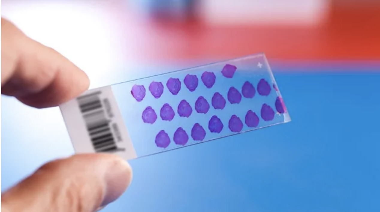
The TMA technique helps to organize small amounts of biological samples. The composite paraffin blocks are constructed by extracting cylindrical core biopsies from different blocks and re-embedding these blocks into a single block with specific array coordinates. The donor blocks are first retrieved, sectioned, and stained with hematoxylin and eosin. After that, an experienced pathologist is required to determine the slides to mark the areas of interest. A TMA equipment can then be used to obtain a core from the donor block. The TMA instrument arraying device produces circular sample spots. This core is then placed at a specific coordinate, which is accurately recorded. This sampling process can then be repeated multiple times until there are hundreds to thousands of cores placed in one recipient block. These are then sectioned using a microtome to generate TMA slides for IHC and molecular analyses. Newer techniques allow as many as 2000 or more sections per slide. With this method, the entire cohort can be analyzed with one or two slides.
Automation
Some have tried to automate the TMA construction process. There are various machines that can array single and multiple TMA blocks in a shorter duration compared to manually. There are also digital techniques that have taken the spotlight in biomedical research and clinical medicine. A high throughput microscopic slide scanner that converts glass slides into digital images have also been developed to allow storage, retrieval, sharing, and algorithmic analysis. The technology that enables the scanning of TMAs into virtual slides will help accelerate the discovery of new biomarkers.
Conclusion
TMA is an effective tool for the analysis of tissues to help identify new markers for diagnostics and prognosis. It also has different degrees of use in research and offers a range of possible applications in research, oncology, and discovery of new therapies. It is thought that TMA will become w widely used tool for all research that uses tissue samples. The TMA technique will also result in significant improvement and advance in the transition of basic research findings to various clinical applications.
References:
1) Jawhar NMT. Tissue microarray: a rapidly evolving diagnostic and research tool. Ann Saudi Med. 2009; 29(2): 123-127.
2) Behling F, Schittenhelm J. Tissue microarrays – translational biomarker research in the fast lane. Expert Review of Molecular Diagnostics. 2018; 18(10): 833-835.


