
Introduction
Tissue microarrays (TMAs) are paraffin blocks where there can be up to 1000 individual tissue cores that are compiled in array fashion and allowing multiplex histological analysis. The TMA technique was developed to address the main limitations in molecular clinical analysis of tissues. The limitations include:
• Complexity of procedures
• Limited availability of diagnostic reagents
• Limited patient size
In 1986, H. Battifora first introduced multi-tissue blocks which were known as “multi-tumor (sausage) tissue block”. This was subsequently modified in 1990 to the “checkerboard tissue block”. J. Kononen and his team then developed the TMA technique in 1998 with a novel sampling approach to produce standardized sizes and shapes allowing it to be precisely arrayed. TMAs contain preserved biomolecules such as carbohydrates, proteins, DNA, RNA, or lipids. this procedure offers a high throughput platform for rapid analysis of molecular markers that are associated with the prognosis or diagnosis of physiological conditions and illnesses. The method is an effective tool with advantages such as:
• Reduction in the workload
• Reduction in assay volume
• Reduction in the number of slides
• Experimental uniformity
• Compatibility with automated staining systems
• Shelf stability up to 12 months when stored at 4°Celsius
Procedure
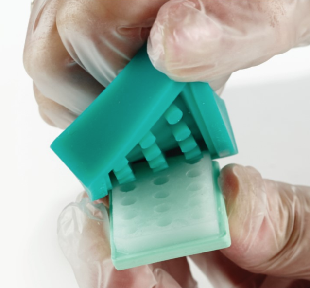
The TMA technique involves using a hollow needle to remove the tissue cores from regions of interest. These tissue cores can be obtained from paraffin-embedded tissues such as tumor samples and clinical biopsies. These tissue cores are inserted in a recipient paraffin block in an array pattern. The block is then sectioned and cut using a microtome, mounted on a microscope, and analyzed using standard histological analysis. Each block can produce about 100 to 500 sections which can be used for various tests. The tests that commonly use TMA sections are fluorescent in situ hybridization (FISH) and immunohistochemistry (IHC). TMAs are also especially useful in the analysis of cancer samples.
Why TMA?
In biology, one of the classic issues would be the validation of a new concept using human tissue. However, it leads to the problem of not having enough tissue in case number or amount to help complete the analysis. The tissue shortage can be attributed to the fact where the initial sampling is performed by a pathologist whose main goal is for diagnostic purposes and not for research. Working with human tissue is also complicated by ethical issues and guidelines where informed consent is required before use. Therefore, once the tissue is obtained, it should be managed properly to maximize its value. TMA is a tool that can help resolve this issue.
For example:
• While conventional slides produce one case per slide, TMA can produce 50 to 500 cases per slide. This maximizes the use of a scarce resource. In conventional slides, 150 sections of 1 case only yields 150 assays while in TMA, 150 sections with more than 500 cases can yield up to about 75,000 assays.
• Conventional slides usually require multiple batches in a single study. Using TMA, the entire cohort can be ready in one experiment.
• Conventional slides need large amounts of reagent while TMA only takes less than 1 ml of total reagent volume for the whole cohort.
• Conventional slides are subject to differential antigen retrieval while TMA is not.
• It is expensive and slow for large cohorts of conventional slides. Using TMA, it is rapid.
TMAs can also be used in various techniques such as immunologic stains (fluorescent visualization or chromogenic), histochemical stains, tissue microdissection, and in situ hybridization (mRNA FISH and in situ hybridization). Even after the tissue has been used for array-based studies, it can still be used to make a diagnosis. This means that the tissue block can be cored several times without destruction of the tissue block.
TMA in Research
TMA combined with IHC is a preferred method to analyze cancer biomarkers in various patient cohorts. The ability to assemble large samples of representative cancer tissue with a corresponding clinical database makes it a powerful resource to learn the correlation between different protein expression patterns and different clinical parameters. Since multiple samples are assembled into one block, the sections can be prepared using the same protocol to help avoid technical artifacts and variability. TMA sets can be used to study cancer biomarkers that play a role in diagnostic, prognostic, and therapy for patients.
Possible Limitations
One of the main limitations of TMA as proposed by experts is the amount of tissue volume being too small and not being able to represent the entire tumor. However, numerous studies have proven that this issue can be compensated by the statistical power of the analysis of hundreds or thousands of cases. Ultimately, the conclusion is not affected as numerous amounts of cases helps to eliminate the variability of a single data point. In more than 95 percent of cases, the staining and analysis of 2 microarray disks are comparable to the analysis of a whole tissue section. The limiting factor is due to the number of sections obtained that depends on the thickness of the donor block. The standard blocks are about 5mm thick. Therefore, when the blocks have been extensively cut and are less than 2.5mm, the array construction is no longer recommended.
References:
1) Tissue microarray. Wikipedia. Accessed 6/25/2019. https://en.wikipedia.org/wiki/Tissue_microarray
2) Tissue microarray. Novus Biologicals. Accessed 6/25/2019. https://www.novusbio.com/product-type/tissue-microarrays
3) Why use tissue microarrays. Yale. Accessed 6/25/2019. https://medicine.yale.edu/pathology/ypts/tissuemicroarrayfacility/why.aspx
