
Tissue microarray (TMA) is a method in pathology that allows researchers to analyze a greater number of tissues than traditional biopsies. Researchers can analyze and test hundreds of tissue samples with customized antibodies for each all at once. This makes tissue microarrays helpful for saving and optimizing time and resources. Here, we’ll discuss the findings of research conducted to apply TMA methods to bone marrow biopsies, the use of TMA for bone marrow biopsies, and the benefits and drawbacks of this new research method.
What is a Tissue Microarray?
TMA’s are typically used to analyze and diagnose cancerous tissue cells. Small tissue samples are collected, embedded with paraffin, and arranged in a paraffin block. A microarray block can include up to 100-500 sections. Each sample can be injected with its antibody, allowing researchers to analyze various samples at once without using excess resources.
Benefits of Tissue Microarray
Paraffin blocks preserve the tissue samples, allowing them to last much longer than other tissue sampling methods. This allows researchers to revisit the samples if need-be and allows them to analyze how they change over an extended period. Tissue microarray is cost-efficient and time-efficient, making it the future of diagnostics and research in the field of medical science and hematopathology (the study of disease and disorders).
Bone Marrow Biopsy (BMB)
Bone marrow biopsies are used to analyze patients who may have trouble creating blood cells. The process of the biopsy involves removing a small amount of bone marrow that exists within your bones. Bone marrow is a soft tissue that is created in the center of large bones, and samples are typically collected from the front of the pelvis.
A pathologist may recommend a bone marrow biopsy if you experience the following:
Anemia (lack of red blood cells)
An abnormal amount of blood cells
Iron deficiency
Cancer present in blood-forming tissue such as leukemia or lymphoma
Cancer that has spread to the bone marrow
Symptoms of chemotherapy
Drawbacks of Bone Marrow Biopsy
Most bone marrow biopsy samples are very small in size due to how invasive they are. This provides health professionals and researchers with a smaller sample to study. A huge advantage of TMA’s is that they provide researchers with sufficient tissue samples. Most bone marrow biopsies are typically no larger than 2.0mm wide and 2.5 cm long. TMA methods, however, require tissue samples of 3-4mm.
According to research conducted by Obermann, Marienhagen, Stoehr, Wuensch, and Hofstaedter, “Availability of TMAs from bone marrow biopsies (BMBs) would be especially useful in hematopathology research because a large number of neoplasms of the lymphatic and hematopoietic system involve the bone marrow. In some entities, such as leukemia, BMBs can be the only type of tissue containing neoplastic cells that is available for the histopathologist.” Therefore, the cost-efficiency and time-efficiency of TMA methods would be incredibly useful for bone marrow biopsies.

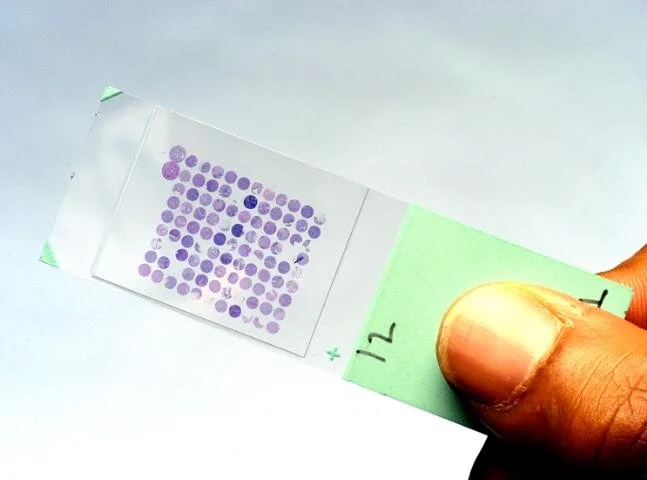
Tissue Microarray constructed from Bone Marrow Biopsies
TMA for Bone Marrow Biopsy Utilizing the Stacking Method
The research previously mentioned adapted tissue microarray methods to better suit bone marrow biopsy samples and meet the specific requirements of BMB. Bone marrow biopsies were selected and inserted into pre-made holes in the TMA block, with a thickness of 0.1 mm and a diameter of 1.6mm. The researchers found that this size optimized the preservation of the biopsy, which is a primary desired advantage of the TMA method.
The Stack Method
The stack method was then used by stacking three punch bone marrow biopsies from one donor block on top of another. The stack method resulted in the final size of the tissue block being 4mm, which is the size required for efficient TMA testing.
Conclusion
After successfully constructing a TMA for a bone marrow biopsy, a total of 100 cells were assessed both in whole sections and tumor cores. The TMA contained 33 cores of 11 BMB’s, and the bone marrow was well preserved - achieving the study’s goal. Tissue microarray is an advantageous method and the possibilities of its application should and can be explored to further our ability to efficiently research, analyze, and diagnose.
Explore our biorepository services and the various cancer types we collect for tissue microarrays.
Resources:
https://www.future-science.com/doi/10.2144/000112073
https://www.jpatholtm.org/journal/Figure.php?xn=kjpathol-46-562.xml&id=
https://en.wikipedia.org/wiki/Tissue_microarray
Limberger, K.A., Bogatyreva, L., Todorova, R. et al. Tissue microarray technique is applicable to bone marrow biopsies of myeloproliferative neoplasms. Histochem Cell Biol 147, 75–82 (2017). https://doi.org/10.1007/s00418-016-1476-x
