Frozen Tissue

Biobanking of Fresh Frozen Tissue from Clinical Surgical Specimens

Fresh Frozen Tissue and Clinical Surgical Specimens


Since many pathology departments at hospitals have procedures for the reception and handling of fresh specimens, a biobanking manual based on the already established structure to enable the collection of unfixed tissue samples can be produced. This allows the collection of all types of surgical lesions. The procedures can be used for all specimens such as a tumor, rejected transplanted organs, atherosclerosis, inflammatory bowel disease, etcetera. Most surgical specimens are transported to the pathology department for potential biobanking. One of the most important concerns for all clinical biobanks is diagnostic security. The pathologist involved has the responsibility to report the diagnosis based on the fresh specimen.

One main limiting factor in frozen tissue biobanking is the well-understood hesitance of pathologists to remove abnormal tissue for biobanking purposes as it may jeopardize the appropriate diagnosis and treatment for the patient. A potential solution to this issue is to perform cryosection and histological examination of the specimen once it enters the biobank. Another important concern for biobanks is the possibility of tissue degradation during transport from the surgical theater to a facility. However, most tissues are usually stable for hours since it is transported on ice. It is also important for each research project to define their tissue quality criteria to ensure that the samples meet their standards.

Tissue Sample Collection and Biobanking


These are some of the methods that can be applied through different stages of biospecimen collection:


1) Surgical Theater

  • Fresh specimen should only be handled in a designated area. Between each case, the area should be decontaminated by removing material from previous cases. Specimens should only be handled using gloves and instruments. Responsibilities of various staff members should be documented.

  • The pathology chart should note the time when the specimen has been removed from the patient.

  • Specimens should be placed in a clean surgical cloth, plastic bag, jars, or a test tube. It should also be immersed in a cold saline solution.

  • The specimen should be transported at ±0⁰C (partly filled with wet ice). It is important to note that the specimen should not be in direct contact with the cooling agent (water or ice) during transport.

  • Inform the technician at the pathology department for reception of the specimen. The communication routine between the theater and pathology department should be safe and clear. Ensure that the specimen is delivered.

2) Pathology Department

  • Upon arrival at the pathology department, the time of arrival should be noted on the chart. It should be registered in the clinical laboratory management system and labeled with a case number.

  • The pathologist on call should be notified regarding the arrival of the specimen. If a delay is inevitable, the specimen should be placed in the refrigerator.

  • Once ready, the specimen is removed and placed on a clean sheet of filter paper. The macroscopic features of the specimen (weight, measurements, description) should be noted in the chart.

  • Pieces of the specimen that represents the lesion and normal tissue should be cut out and placed into a cryomold for cryogel coverage. Storage of samples in cryogel prevents the lyophilization of the specimens. It also helps o keep the DNA and RNA intact. The mold is then snap-freeze in dry ice or isopentane. The time of freezing, biobank numbers, pathologist signature, and technician signatures are noted. These tissue blocks are then transferred to a low-temperature freezer.

  • It should be noted that the thawing of a sample during the lifespan of a fresh frozen biobank sample is one of the most important risk factors for the degradation of tissue. A cryostat to avoid thawing during delivery can be used to slice sections for protein, DNA, or RNA extraction. The tissue lock can also be cracked on a cutting board that has been cooled on dry ice if a larger portion of the sample is needed.

3) Biobank

  • The biobank technician then makes cryosections of the biobank samples.

  • An adhesive tape helps support the section during the cutting and transfer process to prevent folds and tears. Once the section is crossed-linked onto a slide, the tape can be peeled off.

  • Sections are fixed, stained, mounted, and lastly labeled with a case number. These slides are then delivered to the pathologist who is responsible for reporting the diagnosis. The biobank technician registers the case along with relevant information such as name, identification number, age, gender diagnosis, and more.

  • Biobanking protocols concerning biospecimens should be integrated with both local and national established clinical or diagnostic procedures. The protocols should also be authorized by those relevant.


Conclusion

It is critical for research teams involved in molecular diagnostics and translational cancer to have access to quality fresh frozen tissue. This article helps to describe a workflow for the collection of frozen biospecimens after derived from patients after surgery. These routines are used at Uppsala University Hospital since 2001 where the team integrates cryosection and histopathologic examination of the samples in the manual this is to help procure small lesions while avoiding a diagnostic hazard due to the removal of abnormal tissue from the surgical specimen.


References
Botling J, Micke P. Biobanking of fresh frozen tissue from clinical surgical specimens: transport logistics, sample selection, and histologic characterization. Joakim Dillner (ed.). Methods in Biobanking, Methods in Molecular Biology, vol. 675: page 299-306.


Tissue Microarray

Introduction

The recent advances that have occurred in the human molecular genetics field have found that there are gene-based disease mechanisms in various areas of medicine. Studies regarding diagnostic and prognostic markers in many clinical specimens is vital in the translation of new findings from basic science to applications in clinical practice. With the increased use and advancements of new molecular biology methods, the research of progression and pathogenesis of diseases such as cancer are now revolutionized. Understanding the basic molecular mechanisms in the progression of normal tissues to cancerous or malignant tumors is crucial in the knowledge of the disease as it can lead to improved treatment, diagnosis, and cures. Some clinical studies have discovered various novel markers at the gene level where validation of these markers is necessary. However, it can be a time consuming, costly, and labor-intensive process especially if tested on several specimens.


Tissue Microarray

Tissue microarray is a method used in the field of pathology to overcome issues where the validation of markers is:

  • Time-consuming

  • Costly

  • Labor-intensive

It can be used to organize small amounts of tissue samples on a solid support. It is a method designed to allow the:

  • Simultaneous assessment of gene expression on hundreds on tissue samples

  • Parallel molecular profiling of tissue samples at DNA, RNA, and protein level

  • Analysis of samples using fluorescence in situ hybridization (FISH), immunohistochemistry, and RNA in situ hybridization at lower costs and less time


Tissue Microarray Construction

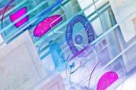
Tissue microarrays can be constructed using composite paraffin blocks through the extraction of cylindrical core biopsies from donor blocks which are them embedded into a microarray or recipient block at specific array coordinates. Donor blocks are first retrieved and sectioned to produce the standard slides. These slides are then stained with hematoxylin and eosin. Once ready, the slides are examined by a certified pathologist who then marks the area of interest (usually an area with pathology such as cancer). Next, the samples can be arrayed. A tissue core can be acquired from the donor block using a tissue microarray instrument. This tissue core is then inserted into an empty recipient or paraffin block at a specific coordinate which is recorded on a spreadsheet. The sampling process is then repeated as many times as necessary from various donor blocks until many cores are placed in one recipient block. This results in the final tissue microarray block. A microtome is utilized to cut 5-micrometer sections from the blocks to produce slides necessary for immunohistochemical and molecular analyses.


Applications and Advantages

Tissue microarrays have many advantages over other techniques. Some of it include:

  1. Amplification of a scarce resource – After a standard histological section that is approximately 3 to 5 millimeters thick is used in primary diagnosis, the sections can further be cut 50 to 100 times yielding a total of 100 assays. In tissue microarrays, instead of 50 to 100 samples, it can produce material enough for 500,000 assays.

  2. Simultaneous analysis – Tissue microarrays allows the simultaneous analysis of many specimens as it provides high throughput data acquisition.

  3. Uniformity – In tissue microarrays, every tissue sample is treated uniformly. It can also be used in a variety of techniques such as fluorescent or chromogenic visualization, histochemical stains, tissue microdissection techniques, and more. Tissue microarray enables the analysis of the entire cohort on one slide standardizing the variables such as incubation times, antigen retrieval, washing procedure, reagent concentration, and temperature.

  4. Time and cost efficient – The tissue microarray method require small amounts of reagents for analysis. It is a method that is both time and cost efficient.

  5. Conservation of tissue samples – Tissue microarray is a technique that does not destroy the original block of the tissue sample.


Tissue Microarrays from Fresh Frozen Tissue

In tissue microarray, the method uses tissue samples from paraffin-embedded tissue donor blocks that are then placed into a recipient block. One of the challenges with paraffin-embedded tissue is the antigenic changes seen in proteins and degradation of mRNA due to the fixation and embedding process. Some researchers have modified the tissue microarray technique by using fresh frozen tissue that is embedded in optimal cutting temperature (OCT) compound. It is then arrayed into a recipient OCT block. Tissue samples are not fixed before the embedding process and the arrayed sections are assessed without fixation. The advantage of tissue microarrays from fresh frozen tissue is that:

  • It works well for DNA, RNA, and protein analyses.

  • Paraffin-embedded tissue arrays can be challenging for RNA in situ hybridization and immunohistochemistry analyses but tissue microarrays from fresh frozen tissue allow the optimal assessment by each technique.

  • It has uniform fixation throughout the whole array panel.

  • It is a technique that may have significant advantages in the assessment of certain genes and proteins as it improves both quantitative and qualitative results.


References:

1)      Jawhar NMT. Tissue microarray: a rapidly evolving diagnostic and research tool. Ann Saudi Med. 2009; 29(2): 123-127.

2)      Fezjo MS, Slamon DJ. Tissue microarrays from frozen tissues – OCT technique. Methods Mol Biol. 2010; 664: 73-80.